Bladder cancer SEM-TEM comparison
Bildnummer 12948538
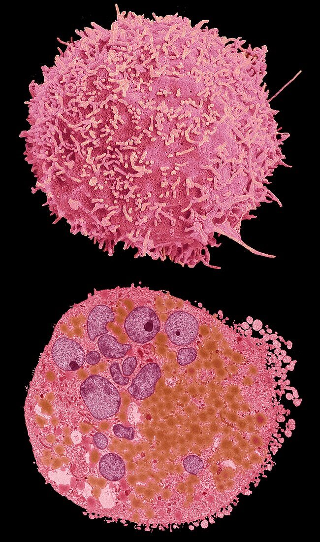
| Comparison between a scanning electron micrograph (SEM, top) and transmission electron micrograph (TEM, bottom) of a bladder cancer cell. Typical of many cancer cells the nucleus (purple in TEM) is enlarged and multilobed, the cell walls are covered in projections (SEM) and the cytoplasm contains a large number of organelles (TEM) including brown lipid droplets. Most bladder cancers arise in the bladder lining. They may spread inwards or through the bladder wall into nearby organs, lymph glands and bones. Smokers and workers in the dye and rubber industries are at increased risk of the disease. It is also more common in tropical areas where the parasitic infection schistosomiasis is prevalent. Symptoms include blood in the urine and bladder infections. Treatment involves excision of affected tissues, with chemotherapy and radiotherapy. Magnification: x3000 when printed at 10cm wide. For a series of comparisons between SEMs and TEMs see images C047/7006 to C047/7034. | |
| Lizenzart: | Lizenzpflichtig |
| Credit: | Science Photo Library / Gschmeissner, Steve |
| Bildgröße: | 4545 px × 7692 px |
| Modell-Rechte: | nicht erforderlich |
| Eigentums-Rechte: | nicht erforderlich |
| Restrictions: | - |
Preise für dieses Bild ab 15 €
Universitäten & Organisationen
(Informationsmaterial Digital, Informationsmaterial Print, Lehrmaterial Digital etc.)
ab 15 €
Redaktionell
(Bücher, Bücher: Sach- und Fachliteratur, Digitale Medien (redaktionell) etc.)
ab 30 €
Werbung
(Anzeigen, Aussenwerbung, Digitale Medien, Fernsehwerbung, Karten, Werbemittel, Zeitschriften etc.)
ab 55 €
Handelsprodukte
(bedruckte Textilie, Kalender, Postkarte, Grußkarte, Verpackung etc.)
ab 75 €
Pauschalpreise
Rechtepakete für die unbeschränkte Bildnutzung in Print oder Online
ab 495 €
Keywords
- abnormal,
- Atomkern,
- Blase,
- farbig,
- gefärbt,
- Gesundheitswesen,
- Kondition,
- Krankheit,
- Krebs,
- krebsartig,
- Lipid,
- maligne,
- Malignom,
- Medizin,
- medizinisch,
- Mikrovilli,
- Organellen,
- rasterelektronenmikroskopische Aufnahme,
- Reihenfolge,
- REM,
- Serie,
- Störung,
- System,
- tem,
- transmissionselektronenmikroskopische Aufnahme,
- Tumor,
- Tumoren,
- Vergleich,
- vergleichen,
- verglichen,
- Wachstum,
- Zelle,
- Zellen