Microvilli, SEM-TEM comparison
Bildnummer 12948534
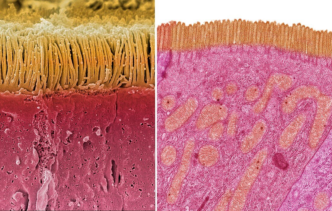
| Intestinal microvilli. Comparison between a scanning electron micrograph (SEM, left) and transmission electron micrograph (TEM, right) of an epithelial cell from a human small intestine, showing the densely packed microvilli. Each microvillus is approximately 1 micrometre long by 0.1 micrometre in diameter and contains a core of actin microfilaments. These tiny structures form a dense brush-like covering on the absorptive surfaces of the cells lining the small intestine. The cells absorb nutrients from digested food through the microvilli. Orange mitochondria are visible in the cytoplasm of the cell in the TEM image. Magnification: x3000 when printed at 10 centimetres wide. For a series of comparisons between SEMs and TEMs see images C047/7006 to C047/7034. | |
| Lizenzart: | Lizenzpflichtig |
| Credit: | Science Photo Library / Gschmeissner, Steve |
| Bildgröße: | 7180 px × 4572 px |
| Modell-Rechte: | nicht erforderlich |
| Eigentums-Rechte: | nicht erforderlich |
| Restrictions: | - |
Preise für dieses Bild ab 15 €
Universitäten & Organisationen
(Informationsmaterial Digital, Informationsmaterial Print, Lehrmaterial Digital etc.)
ab 15 €
Redaktionell
(Bücher, Bücher: Sach- und Fachliteratur, Digitale Medien (redaktionell) etc.)
ab 30 €
Werbung
(Anzeigen, Aussenwerbung, Digitale Medien, Fernsehwerbung, Karten, Werbemittel, Zeitschriften etc.)
ab 55 €
Handelsprodukte
(bedruckte Textilie, Kalender, Postkarte, Grußkarte, Verpackung etc.)
ab 75 €
Pauschalpreise
Rechtepakete für die unbeschränkte Bildnutzung in Print oder Online
ab 495 €
Keywords
- Aktin,
- Biologie,
- biologisch,
- Darm,
- Farbig,
- gefärbt,
- gesund,
- Kultur,
- menschlicher Körper,
- Mikroskop,
- Mikrovilli,
- Mitochondrien,
- normal,
- rasterelektronenmikroskopische Aufnahme,
- Reihenfolge,
- REM,
- Serie,
- Struktur,
- tem,
- Transmissionselektronenmikroskopie,
- transmissionselektronenmikroskopische Aufnahme,
- Vergleich,
- vergleichen,
- verglichen,
- Wimpern,
- Zelle