Skin, SEM-TEM comparison
Bildnummer 12948524
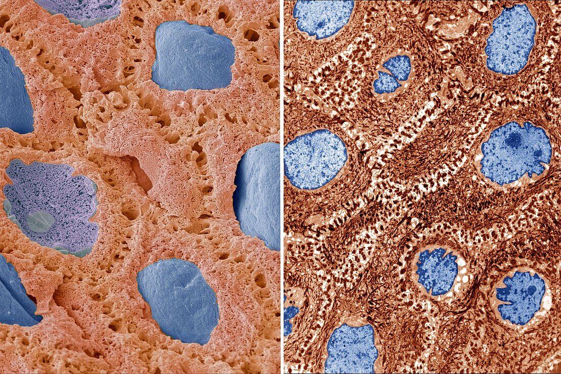
| Skin. Comparison between a scanning electron micrograph (SEM, left) and transmission electron micrograph (TEM, right) of keratinocyte skin cells, which are found in the epidermis layer of the skin. The cells have a large nucleus (blue), which contains their genetic information. In the TEM image the cells' cytoplasm is rich in filaments of the protein cytokeratin (dark brown). The filaments extend into junctions between the cells known as desmosomes, which help the cells to withstand mechanical stress. Magnification: x4000 when printed 10 centimetres high.. For a series of comparisons between SEMs and TEMs see images C047/7006 to C047/7034. | |
| Lizenzart: | Lizenzpflichtig |
| Credit: | Science Photo Library / Gschmeissner, Steve |
| Bildgröße: | 6845 px × 4569 px |
| Modell-Rechte: | nicht erforderlich |
| Eigentums-Rechte: | nicht erforderlich |
| Restrictions: | - |
Preise für dieses Bild ab 15 €
Universitäten & Organisationen
(Informationsmaterial Digital, Informationsmaterial Print, Lehrmaterial Digital etc.)
ab 15 €
Redaktionell
(Bücher, Bücher: Sach- und Fachliteratur, Digitale Medien (redaktionell) etc.)
ab 30 €
Werbung
(Anzeigen, Aussenwerbung, Digitale Medien, Fernsehwerbung, Karten, Werbemittel, Zeitschriften etc.)
ab 55 €
Handelsprodukte
(bedruckte Textilie, Kalender, Postkarte, Grußkarte, Verpackung etc.)
ab 75 €
Pauschalpreise
Rechtepakete für die unbeschränkte Bildnutzung in Print oder Online
ab 495 €
Keywords
- Anatomie,
- anatomisch,
- Atomkern,
- Biologie,
- biologisch,
- epidermal,
- Epidermis,
- farbig,
- Filament,
- Filamente,
- gefärbt,
- Haut,
- Keratinozyten,
- Kerne,
- menschlicher Körper,
- Oberfläche,
- rasterelektronenmikroskopische Aufnahme,
- Reihenfolge,
- REM,
- Serie,
- tem,
- Transmissionselektronenmikroskop,
- transmissionselektronenmikroskopische Aufnahme,
- Vergleich,
- vergleichen,
- verglichen,
- Zelle,
- Zellen