Recurrent glioma brain tumour, 3D and 2D MRI scans
Bildnummer 12948285
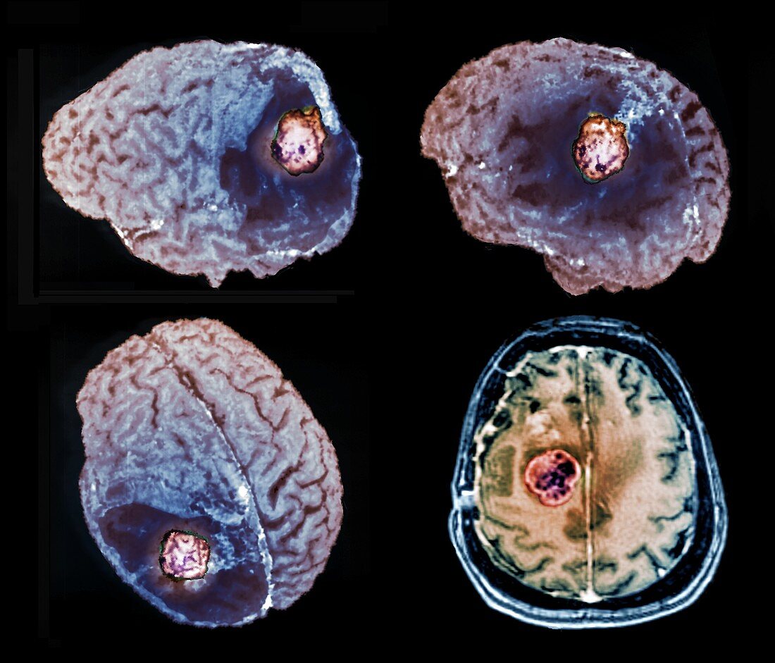
| Recurrent glioma brain tumour. Coloured 3D and 2D magnetic resonance imaging (MRI) scans of the brain of a 60-year-old man who was operated on for a glioma brain tumour which has recurred (coloured mass). An axial scan is at lower right. This malignant tumour (cancer) arises from a type of glial (support) cell of the brain. The cancer is in the brain's left hemisphere and has returned 2 years after the initial operation. This cancer does not spread, but will damage the brain if not treated by surgery, radiotherapy or anti-cancer drugs. | |
| Lizenzart: | Lizenzpflichtig |
| Credit: | Science Photo Library / Zephyr |
| Bildgröße: | 3969 px × 3396 px |
| Modell-Rechte: | nicht erforderlich |
| Eigentums-Rechte: | nicht erforderlich |
| Restrictions: | - |
Preise für dieses Bild ab 15 €
Universitäten & Organisationen
(Informationsmaterial Digital, Informationsmaterial Print, Lehrmaterial Digital etc.)
ab 15 €
Redaktionell
(Bücher, Bücher: Sach- und Fachliteratur, Digitale Medien (redaktionell) etc.)
ab 30 €
Werbung
(Anzeigen, Aussenwerbung, Digitale Medien, Fernsehwerbung, Karten, Werbemittel, Zeitschriften etc.)
ab 55 €
Handelsprodukte
(bedruckte Textilie, Kalender, Postkarte, Grußkarte, Verpackung etc.)
ab 75 €
Pauschalpreise
Rechtepakete für die unbeschränkte Bildnutzung in Print oder Online
ab 495 €
Keywords
- 3-d,
- 3D,
- 60er Jahre,
- ausgeschnitten,
- Ausschnitte,
- axial,
- Diagnose,
- Dreidimensional,
- Erwachsene,
- farbig,
- geduldig,
- gefärbt,
- Gehirn,
- Gliom,
- Hirntumor,
- Kondition,
- Kopf,
- Krankheit,
- Krebs,
- krebsartig,
- linke Hemisphäre,
- Magnetresonanztomografie,
- maligne,
- Malignom,
- Mann,
- Männlich,
- Medizin,
- medizinisch,
- menschlicher Körper,
- MRT-Untersuchung,
- neural,
- Neurologie,
- neurologisch,
- Niemand,
- Onkologie,
- Quartett,
- Reihenfolge,
- Scanner,
- schwarzer Hintergrund,
- sechziger Jahre,
- Serie,
- Störung,
- Tumor,
- vier,
- Wachstum,
- Wiederkehrend