Vascular disease,CT scans
Bildnummer 12896634
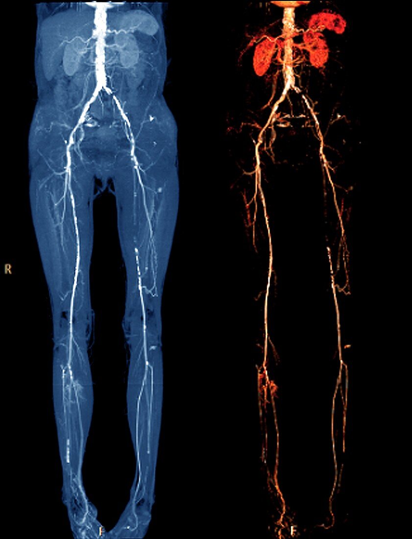
| Vascular disease. Coloured 2D (left) and 3D (right) computed tomography (CT) angiograms of the abdominal aorta and lower limbs of a 68-year-old woman with vascular disease,showing the presence of diffuse calcified atheromas,an occlusion of the left iliac artery and left hypogastric artery,and stenosis and infiltration of the femoral arteries and tibioperoneal arteries. | |
| Lizenzart: | Lizenzpflichtig |
| Credit: | Science Photo Library / Zephyr |
| Bildgröße: | 3402 px × 4448 px |
| Modell-Rechte: | nicht erforderlich |
| Eigentums-Rechte: | nicht erforderlich |
| Restrictions: | - |
Preise für dieses Bild ab 15 €
Universitäten & Organisationen
(Informationsmaterial Digital, Informationsmaterial Print, Lehrmaterial Digital etc.)
ab 15 €
Redaktionell
(Bücher, Bücher: Sach- und Fachliteratur, Digitale Medien (redaktionell) etc.)
ab 30 €
Werbung
(Anzeigen, Aussenwerbung, Digitale Medien, Fernsehwerbung, Karten, Werbemittel, Zeitschriften etc.)
ab 55 €
Handelsprodukte
(bedruckte Textilie, Kalender, Postkarte, Grußkarte, Verpackung etc.)
ab 75 €
Pauschalpreise
Rechtepakete für die unbeschränkte Bildnutzung in Print oder Online
ab 495 €
Keywords
- 3D,
- 60er Jahre,
- abnormal,
- Angiogramm,
- Aorta,
- Arterie,
- arteriell,
- Arterien,
- Atherom,
- Atherosklerose,
- Bauch,
- Bein,
- Beine,
- Blutgefäß,
- Computertomographie,
- CT-Scan,
- diagnostische Bildgebung,
- Dreidimensional,
- farbig,
- Femoralarterie,
- gefärbt,
- Gefäße,
- Gefässkrankheit,
- Gesundheitswesen,
- Glied,
- Kondition,
- krank,
- Kreislauf,
- Medizin,
- medizinisch,
- menschlicher Körper,
- Niemand,
- peripher,
- Plague,
- Radiographie,
- Radiologie,
- radiologisch,
- Röntgen,
- Röntgenstrahlen,
- Röntgenstrahlung,
- schwarzer Hintergrund,
- sechziger Jahre,
- Störung,
- ungesund