Second Trimester scan for Spina Bifida
Bildnummer 12652581
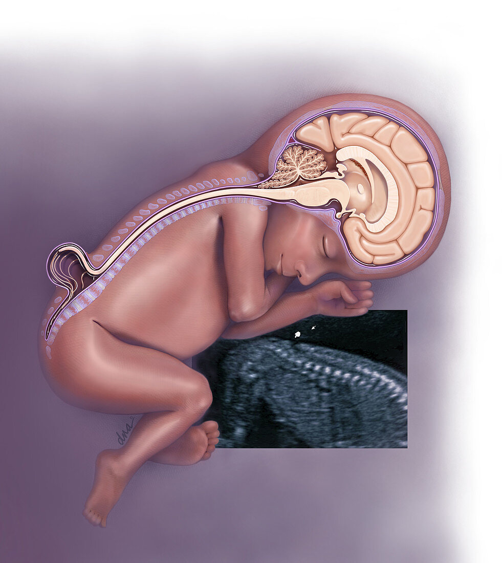
| Illustration to accompany article on use of ultrasound to identify majority of major structural fetal anomalies. Shown is a scan on a 20 week foetus with Spina Bifida. The base of the brain has a Arnold-Chiari Type II malformation (herniation of the hindbrain into the spine). There is thinning of the corpus callosum from ventriculomegaly. The lower spine is shown with a myelomeninocele. | |
| Lizenzart: | Lizenzpflichtig |
| Credit: | Science Photo Library / Science Source / DNA Illustrations |
| Bildgröße: | 3013 px × 3379 px |
| Modell-Rechte: | nicht erforderlich |
| Eigentums-Rechte: | nicht erforderlich |
| Restrictions: | - |
Preise für dieses Bild ab 15 €
Universitäten & Organisationen
(Informationsmaterial Digital, Informationsmaterial Print, Lehrmaterial Digital etc.)
ab 15 €
Redaktionell
(Bücher, Bücher: Sach- und Fachliteratur, Digitale Medien (redaktionell) etc.)
ab 30 €
Werbung
(Anzeigen, Aussenwerbung, Digitale Medien, Fernsehwerbung, Karten, Werbemittel, Zeitschriften etc.)
ab 55 €
Handelsprodukte
(bedruckte Textilie, Kalender, Postkarte, Grußkarte, Verpackung etc.)
ab 75 €
Pauschalpreise
Rechtepakete für die unbeschränkte Bildnutzung in Print oder Online
ab 495 €