Innervation of Cochlea and Ear Canal, Illustration
Bildnummer 12651164
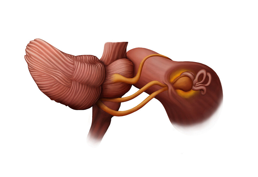
| 2D illustration of the nerves (shown in gold) that connect the brain to the ear canal and cochlea. The cerebellum, brainstem, and cochlea are all shown in relation to these nerves at a posterolateral view. The trigeminal nerve (top) stems from the pons of the brainstem and branches out across the face to provide sensation within this part of the body. The facial nerve (middle) provides sensation to multiple areas around the head, such as the external auditory canal, facial muscles, and part of the tongue. The vestibulocochlear nerve (bottom) stretches from the pons to the inner ear, and helps to send messages from the inner ear to the brain, makes hearing possible, and is essential for balance. | |
| Lizenzart: | Lizenzpflichtig |
| Credit: | Science Photo Library / Science Source / Emily Ciosek |
| Bildgröße: | 3102 px × 2123 px |
| Modell-Rechte: | nicht erforderlich |
| Eigentums-Rechte: | nicht erforderlich |
| Restrictions: | - |
Preise für dieses Bild ab 15 €
Universitäten & Organisationen
(Informationsmaterial Digital, Informationsmaterial Print, Lehrmaterial Digital etc.)
ab 15 €
Redaktionell
(Bücher, Bücher: Sach- und Fachliteratur, Digitale Medien (redaktionell) etc.)
ab 30 €
Werbung
(Anzeigen, Aussenwerbung, Digitale Medien, Fernsehwerbung, Karten, Werbemittel, Zeitschriften etc.)
ab 55 €
Handelsprodukte
(bedruckte Textilie, Kalender, Postkarte, Grußkarte, Verpackung etc.)
ab 75 €
Pauschalpreise
Rechtepakete für die unbeschränkte Bildnutzung in Print oder Online
ab 495 €