Embryo and Neural Tube
Bildnummer 12650237
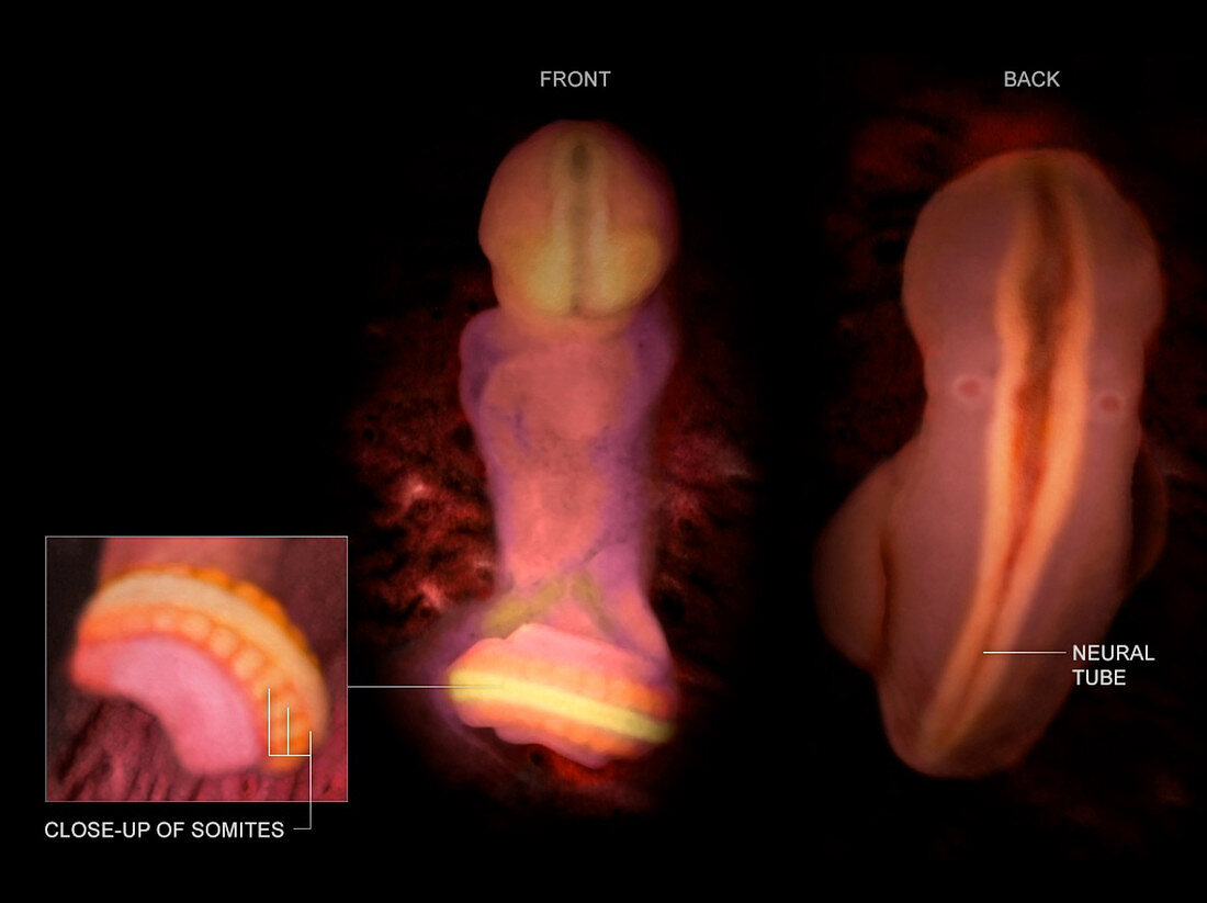
| Medical visualization taken from human scanned data showing an anterior and posterior view of an embryo at week 6 after fertilization. An inset is also shown that displays the somites (ridges on the tail that are the foundation of the skeletomuscular system) in more detail. In the front view, the head (top) can be seen bending downward. The tubelike form in the head is the neural tube, formed by the closing of the neural groove 28 days after fertilization. When the tube doesn't close completely, neural tube defects (NTDs) develop. NTDs are common birth defects, occurring in about 1 in 1, 000 live births. | |
| Lizenzart: | Lizenzpflichtig |
| Credit: | Science Photo Library / Science Source / TheVisualMD |
| Bildgröße: | 2673 px × 2000 px |
| Modell-Rechte: | nicht erforderlich |
| Eigentums-Rechte: | nicht erforderlich |
| Restrictions: | - |
Preise für dieses Bild ab 15 €
Universitäten & Organisationen
(Informationsmaterial Digital, Informationsmaterial Print, Lehrmaterial Digital etc.)
ab 15 €
Redaktionell
(Bücher, Bücher: Sach- und Fachliteratur, Digitale Medien (redaktionell) etc.)
ab 30 €
Werbung
(Anzeigen, Aussenwerbung, Digitale Medien, Fernsehwerbung, Karten, Werbemittel, Zeitschriften etc.)
ab 55 €
Handelsprodukte
(bedruckte Textilie, Kalender, Postkarte, Grußkarte, Verpackung etc.)
ab 75 €
Pauschalpreise
Rechtepakete für die unbeschränkte Bildnutzung in Print oder Online
ab 495 €