Aorta, Cross Section, SEM
Bildnummer 12649042
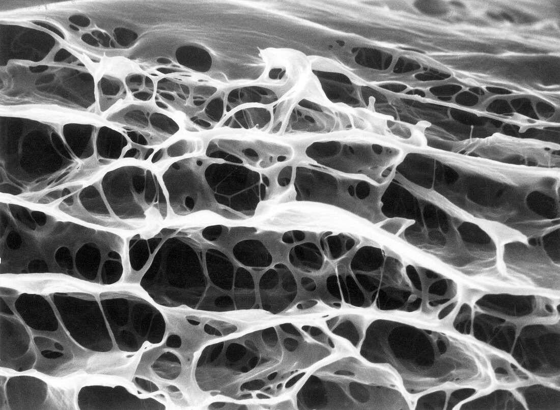
| Scanning electron micrograph of the wall of an aorta in cross-section, revealing the architecture of the elastin in 3-D. Formic acid has removed all other tissue components. The elastic tissue consists of multiple concentric sheets, or laminae, interconnected by radially oriented strands and fenestrated septa. In an intact vessel, smooth muscle cells occupy the spaces demarcated by the elastin elements. SEM, magnification 2400x. | |
| Lizenzart: | Lizenzpflichtig |
| Credit: | Science Photo Library / Science Source / Don W. Fawcett |
| Bildgröße: | 3866 px × 2834 px |
| Modell-Rechte: | nicht erforderlich |
| Eigentums-Rechte: | nicht erforderlich |
| Restrictions: | - |
Preise für dieses Bild ab 15 €
Universitäten & Organisationen
(Informationsmaterial Digital, Informationsmaterial Print, Lehrmaterial Digital etc.)
ab 15 €
Redaktionell
(Bücher, Bücher: Sach- und Fachliteratur, Digitale Medien (redaktionell) etc.)
ab 30 €
Werbung
(Anzeigen, Aussenwerbung, Digitale Medien, Fernsehwerbung, Karten, Werbemittel, Zeitschriften etc.)
ab 55 €
Handelsprodukte
(bedruckte Textilie, Kalender, Postkarte, Grußkarte, Verpackung etc.)
ab 75 €
Pauschalpreise
Rechtepakete für die unbeschränkte Bildnutzung in Print oder Online
ab 495 €