Blood Vessel, SEM
Bildnummer 12649037
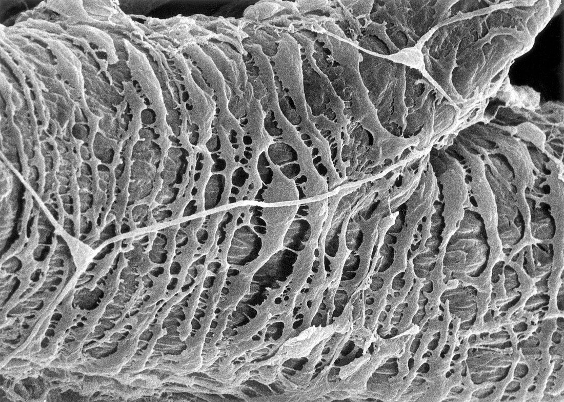
| Scanning electron micrograph of a blood vessel. A large venule is joined by a smaller tributary at upper right. Smooth muscle cells are more irregular in shape and are spaced farther apart than arterioles. Elements of the perivascular nerve net can be seen branching over the vessel. | |
| Lizenzart: | Lizenzpflichtig |
| Credit: | Science Photo Library / Science Source / Don W. Fawcett |
| Bildgröße: | 4026 px × 2871 px |
| Modell-Rechte: | nicht erforderlich |
| Eigentums-Rechte: | nicht erforderlich |
| Restrictions: | - |
Preise für dieses Bild ab 15 €
Universitäten & Organisationen
(Informationsmaterial Digital, Informationsmaterial Print, Lehrmaterial Digital etc.)
ab 15 €
Redaktionell
(Bücher, Bücher: Sach- und Fachliteratur, Digitale Medien (redaktionell) etc.)
ab 30 €
Werbung
(Anzeigen, Aussenwerbung, Digitale Medien, Fernsehwerbung, Karten, Werbemittel, Zeitschriften etc.)
ab 55 €
Handelsprodukte
(bedruckte Textilie, Kalender, Postkarte, Grußkarte, Verpackung etc.)
ab 75 €
Pauschalpreise
Rechtepakete für die unbeschränkte Bildnutzung in Print oder Online
ab 495 €