MRI of Arterial Venous Fistulas 5
Bildnummer 12648190
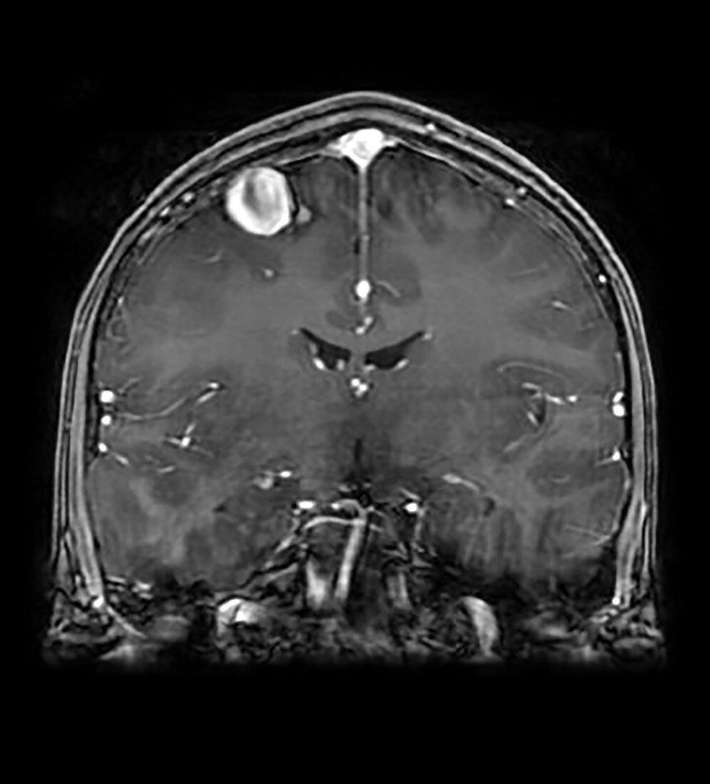
| This coronal (frontal view) T1 weighted MR image with contrast shows an enlarged vascular channel along the surface of the frontal lobe on viewers left reflecting an abnormal connection between an artery and vein (with abscess of the intervening capillary bed). This is called an AV (arterial venous) fistula. There is also enlargement of the anterior cerebral artery secondary to a second AV fistula. | |
| Lizenzart: | Lizenzpflichtig |
| Credit: | Science Photo Library / Science Source / Living Art Enterprises, LLC |
| Bildgröße: | 3900 px × 4307 px |
| Modell-Rechte: | nicht erforderlich |
| Eigentums-Rechte: | nicht erforderlich |
| Restrictions: | - |
Preise für dieses Bild ab 15 €
Universitäten & Organisationen
(Informationsmaterial Digital, Informationsmaterial Print, Lehrmaterial Digital etc.)
ab 15 €
Redaktionell
(Bücher, Bücher: Sach- und Fachliteratur, Digitale Medien (redaktionell) etc.)
ab 30 €
Werbung
(Anzeigen, Aussenwerbung, Digitale Medien, Fernsehwerbung, Karten, Werbemittel, Zeitschriften etc.)
ab 55 €
Handelsprodukte
(bedruckte Textilie, Kalender, Postkarte, Grußkarte, Verpackung etc.)
ab 75 €
Pauschalpreise
Rechtepakete für die unbeschränkte Bildnutzung in Print oder Online
ab 495 €