Epidermoid Tumour on MRI
Bildnummer 12647026
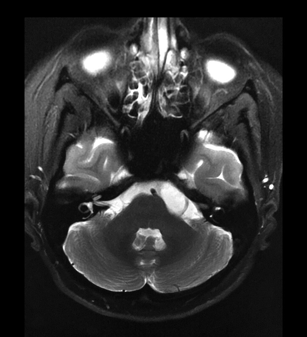
| This axial (cross sectional) T2 weighted non-contrast image of the brain shows subtle deformity of the pre-ganglionic segment of the left trigeminal nerve and mild asymmetric widening of the cerebello-pontine angle cister (CP angle) on viewers right. This is secondary to the presence of a congenital epidermoid tumour which consists of desquamated squamous epithelium with keratin debri. These are benign but can increase in size. They often encase adjacent nerves and blood vessels which can make it difficult to remove. | |
| Lizenzart: | Lizenzpflichtig |
| Credit: | Science Photo Library / Science Source / Living Art Enterprises, LLC |
| Bildgröße: | 3900 px × 4292 px |
| Modell-Rechte: | nicht erforderlich |
| Eigentums-Rechte: | nicht erforderlich |
| Restrictions: | - |
Preise für dieses Bild ab 15 €
Universitäten & Organisationen
(Informationsmaterial Digital, Informationsmaterial Print, Lehrmaterial Digital etc.)
ab 15 €
Redaktionell
(Bücher, Bücher: Sach- und Fachliteratur, Digitale Medien (redaktionell) etc.)
ab 30 €
Werbung
(Anzeigen, Aussenwerbung, Digitale Medien, Fernsehwerbung, Karten, Werbemittel, Zeitschriften etc.)
ab 55 €
Handelsprodukte
(bedruckte Textilie, Kalender, Postkarte, Grußkarte, Verpackung etc.)
ab 75 €
Pauschalpreise
Rechtepakete für die unbeschränkte Bildnutzung in Print oder Online
ab 495 €