Foetal skeletons diorama, 17th century
Bildnummer 12646141
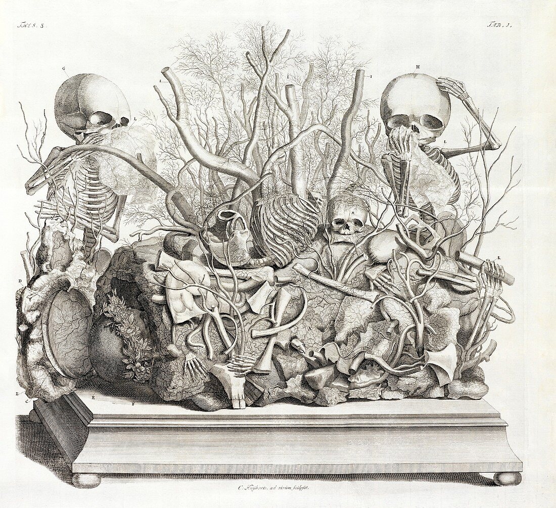
| Foetal skeletons diorama. 18th-century illustration of one of the foetal skeleton dioramas assembled by Dutch anatomist Frederik Ruysch (1638-1731), which he put on display to the public at his museum ('cabinet of curiosities') in Amsterdam towards the end of the 17th century. This example includes foetal and infant skeletons, arrayed with internal organs and tissues from human bodies. These include intestines, dried arteries and veins, and gallstones and kidney stones. Two of the skeletons are shown wiping tears away with 'handkerchiefs'. These illustrations of the dioramas were published from 1701 to 1716 as engravings by Cornelius Huyberts in 'Thesaurus anatomicus primus'. | |
| Lizenzart: | Lizenzpflichtig |
| Credit: | Science Photo Library / THE GETTY |
| Bildgröße: | 4735 px × 4324 px |
| Modell-Rechte: | nicht erforderlich |
| Eigentums-Rechte: | nicht erforderlich |
| Restrictions: | - |
Preise für dieses Bild ab 15 €
Universitäten & Organisationen
(Informationsmaterial Digital, Informationsmaterial Print, Lehrmaterial Digital etc.)
ab 15 €
Redaktionell
(Bücher, Bücher: Sach- und Fachliteratur, Digitale Medien (redaktionell) etc.)
ab 30 €
Werbung
(Anzeigen, Aussenwerbung, Digitale Medien, Fernsehwerbung, Karten, Werbemittel, Zeitschriften etc.)
ab 55 €
Handelsprodukte
(bedruckte Textilie, Kalender, Postkarte, Grußkarte, Verpackung etc.)
ab 75 €
Pauschalpreise
Rechtepakete für die unbeschränkte Bildnutzung in Print oder Online
ab 495 €
Keywords
- 1600er Jahre,
- 17. Jahrhundert,
- 1700er Jahre,
- 18. Jahrhundert,
- Abstrakt,
- Amsterdam,
- Anatomie,
- anatomisch,
- Einfarbig,
- Europa,
- europäisch,
- Geschichte,
- historisch,
- Illustration,
- Konzept,
- Konzepte,
- konzeptionell,
- Künstlerisch,
- Kunstwerk,
- Medizin,
- medizinisch,
- menschlicher Körper,
- Morbid,
- Museum,
- Neugierde,
- Niederlande,
- Niederländisch,
- Niemand,
- Schicksal,
- Schwarz und weiß,
- Skelett,
- symbolisch,
- Tod,
- Tot