Aortic valve replacement, 2D and 3D CT scans
Bildnummer 12644567
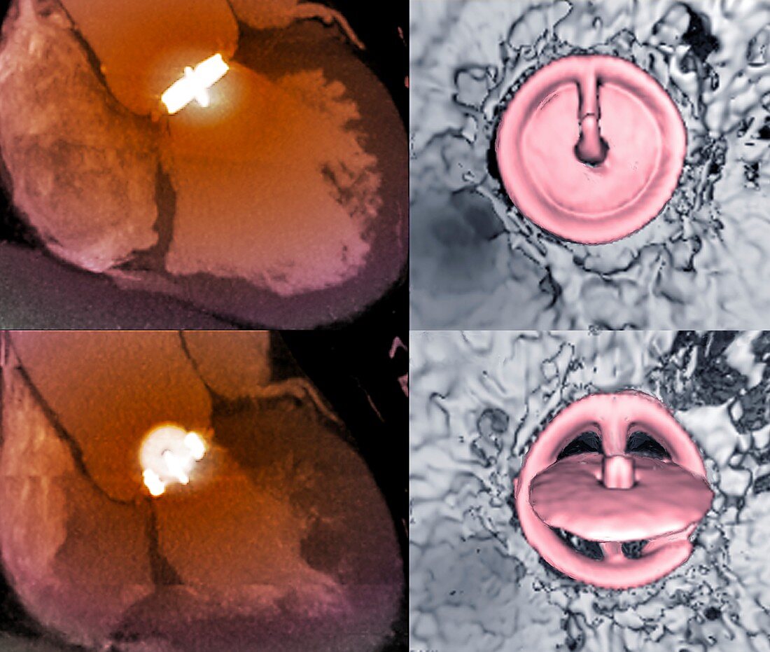
| Aortic valve replacement. Coloured 2D (left) and 3D (right) computed tomography (CT) scans of a replacement aortic valve in the heart of a 58-year-old woman who was being treated for cardiac insufficiency. The aortic valve is shown both in its open (systolic) position (across bottom) and in its closed (diastolic) position (across top). This artificial valve is a Medtronic-Hall valve, consisting of a pyrolytic carbon disc oscillating around an eccentric axis inside a ring. The aortic valve lies between the heart's left ventricle and the aorta. | |
| Lizenzart: | Lizenzpflichtig |
| Credit: | Science Photo Library / Zephyr |
| Bildgröße: | 4544 px × 3846 px |
| Modell-Rechte: | nicht erforderlich |
| Eigentums-Rechte: | nicht erforderlich |
| Restrictions: | - |
Preise für dieses Bild ab 15 €
Universitäten & Organisationen
(Informationsmaterial Digital, Informationsmaterial Print, Lehrmaterial Digital etc.)
ab 15 €
Redaktionell
(Bücher, Bücher: Sach- und Fachliteratur, Digitale Medien (redaktionell) etc.)
ab 30 €
Werbung
(Anzeigen, Aussenwerbung, Digitale Medien, Fernsehwerbung, Karten, Werbemittel, Zeitschriften etc.)
ab 55 €
Handelsprodukte
(bedruckte Textilie, Kalender, Postkarte, Grußkarte, Verpackung etc.)
ab 75 €
Pauschalpreise
Rechtepakete für die unbeschränkte Bildnutzung in Print oder Online
ab 495 €
Keywords
- 3 dimensional,
- 3-d,
- 3-dimensional,
- 3D,
- 50er Jahre,
- Aorta,
- behandelt,
- chirurgisch,
- Computertomographie,
- CT-Scan,
- Dreidimensional,
- Ersatz,
- Erwachsene,
- farbig,
- Fünfziger Jahre,
- geduldig,
- gefärbt,
- Herz,
- Implantat,
- Kardiologie,
- kardiovaskular,
- Kondition,
- Kreislauf,
- Mann,
- Männlich,
- Medizin,
- medizinisch,
- menschlicher Körper,
- Niemand,
- Öffnen,
- Operation,
- Prothese,
- Quartett,
- repariert,
- Scanner,
- Störung,
- Technologie,
- technologisch,
- Ventil,
- vier