Heart and lungs, CT scan
Bildnummer 12644347
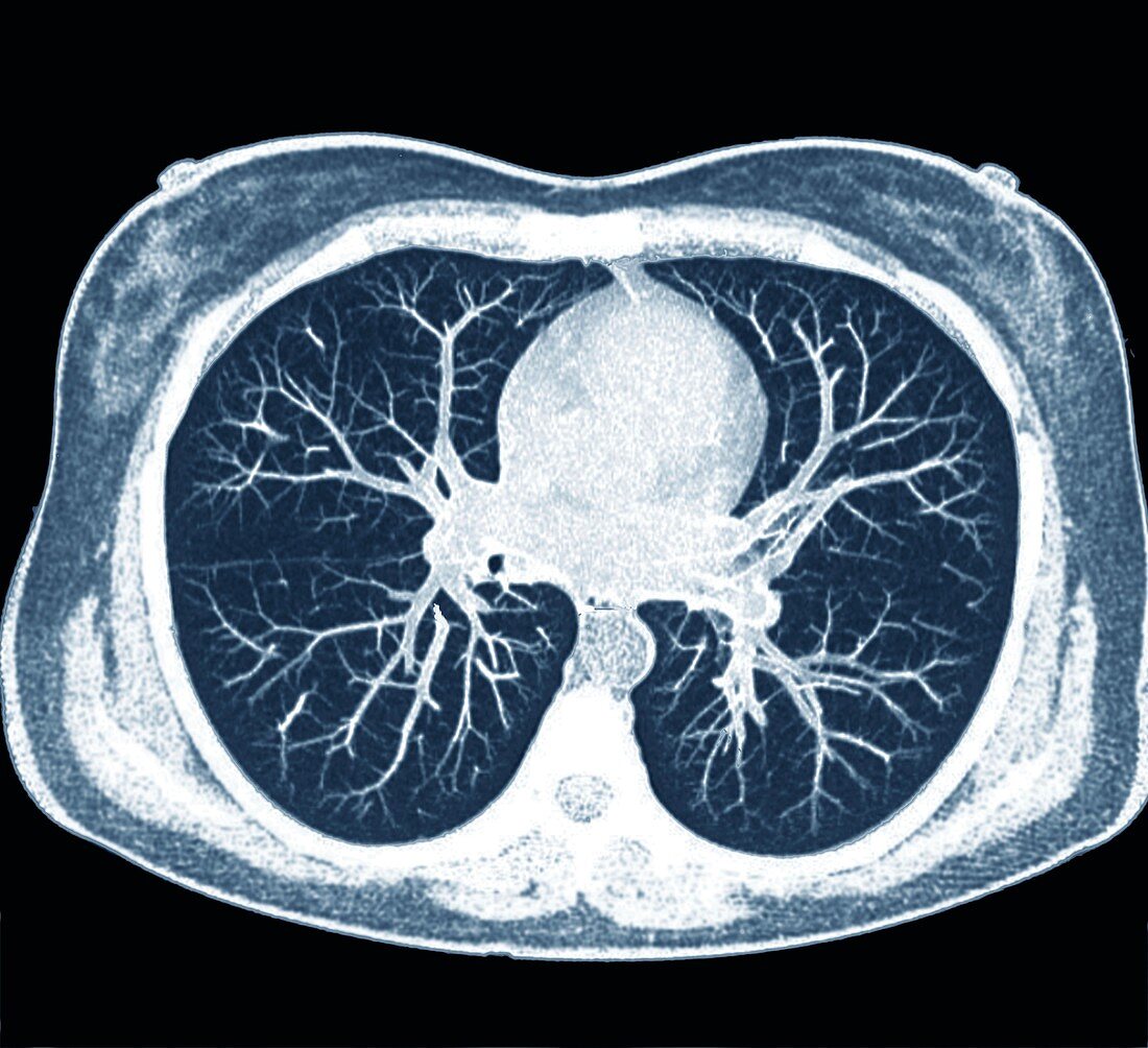
| Heart and lungs. Axial computed tomography (CT) scan of the chest of a 42-year-old woman, showing the normal heart and lungs as viewed from below. The heart (upper centre) is located between the two lungs (left and right). Blood vessels and airways are seen branching out in the lungs. Bone structures include the ribs, the breastbone (sternum) and the backbone (spine). | |
| Lizenzart: | Lizenzpflichtig |
| Credit: | Science Photo Library / Zephyr |
| Bildgröße: | 4375 px × 3995 px |
| Modell-Rechte: | nicht erforderlich |
| Eigentums-Rechte: | nicht erforderlich |
| Restrictions: | - |
Preise für dieses Bild ab 15 €
Universitäten & Organisationen
(Informationsmaterial Digital, Informationsmaterial Print, Lehrmaterial Digital etc.)
ab 15 €
Redaktionell
(Bücher, Bücher: Sach- und Fachliteratur, Digitale Medien (redaktionell) etc.)
ab 30 €
Werbung
(Anzeigen, Aussenwerbung, Digitale Medien, Fernsehwerbung, Karten, Werbemittel, Zeitschriften etc.)
ab 55 €
Handelsprodukte
(bedruckte Textilie, Kalender, Postkarte, Grußkarte, Verpackung etc.)
ab 75 €
Pauschalpreise
Rechtepakete für die unbeschränkte Bildnutzung in Print oder Online
ab 495 €
Keywords
- 40er Jahre,
- Anatomie,
- anatomisch,
- Atemwege,
- ausgeschnitten,
- Ausschnitte,
- axial,
- Biologie,
- biologisch,
- Blau,
- Blutgefäße,
- Computertomographie,
- CT-Scan,
- Einfarbig,
- Erwachsene,
- Frau,
- gesund,
- Herz,
- kardiovaskular,
- Kreislauf,
- Lunge,
- Lungen,
- menschlicher Körper,
- Niemand,
- normal,
- Organ,
- pulmonal,
- Querschnitt,
- Scanner,
- schwarzer Hintergrund,
- Sektion,
- sektioniert,
- Thorax,
- Truhe,
- vaskulär,
- Vierziger Jahre,
- Weiblich