Tongue cancer, CT scan
Bildnummer 12641630
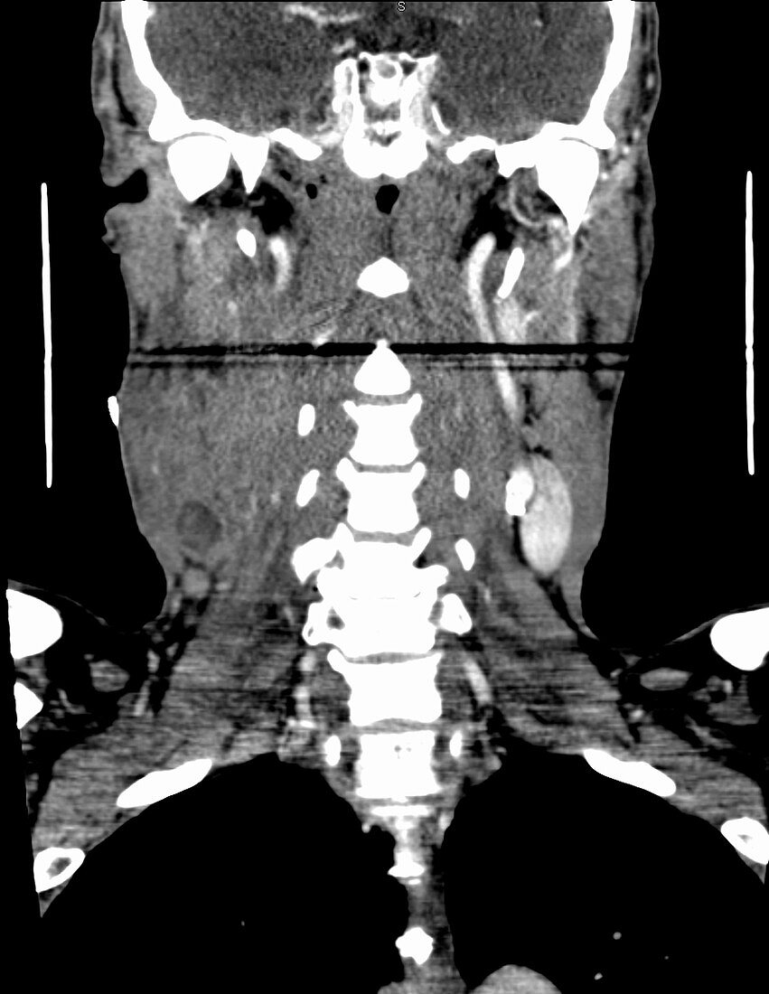
| 60 year old male with right squamous cell carcinoma of the tongue. Volume CT scan of the neck with contrast. 2.5 mm coronal image shows a large confluent indistinctly marginated soft tissue mass along the right side of the neck, extending from the angle of the mandible inferiorly to the level of the thyroid cartilage, approximately 7.8 cm CC x 5.3 cm AP x 4.5 cm transverse. Laterally, the mass infiltrates and enlarges the sternocleidomastoid muscle. Medially, the mass extends to the level of the hypopharynx and completely encases the internal and external carotid artery on the right without appreciable narrowing of those vessels. The mass is predominately isodense to muscle with a 1 cm low attenuation component inferiorly consistent with necrosis or haemorrhage. The confluent lymphadenopathy may be seen with lymphoma and metastatic nasopharyngeal carcinoma. | |
| Lizenzart: | Lizenzpflichtig |
| Credit: | Science Photo Library / Steven Needell |
| Bildgröße: | 1764 px × 2280 px |
| Modell-Rechte: | nicht erforderlich |
| Eigentums-Rechte: | nicht erforderlich |
| Restrictions: | - |
Preise für dieses Bild ab 15 €
Universitäten & Organisationen
(Informationsmaterial Digital, Informationsmaterial Print, Lehrmaterial Digital etc.)
ab 15 €
Redaktionell
(Bücher, Bücher: Sach- und Fachliteratur, Digitale Medien (redaktionell) etc.)
ab 30 €
Werbung
(Anzeigen, Aussenwerbung, Digitale Medien, Fernsehwerbung, Karten, Werbemittel, Zeitschriften etc.)
ab 55 €
Handelsprodukte
(bedruckte Textilie, Kalender, Postkarte, Grußkarte, Verpackung etc.)
ab 75 €
Pauschalpreise
Rechtepakete für die unbeschränkte Bildnutzung in Print oder Online
ab 495 €
Keywords
- abnormal,
- Arterie,
- Ausbreitung,
- axial,
- Bildgebung,
- Blutung,
- Computertomographie,
- ct,
- CT-Scan,
- Diagnose,
- Hals,
- Karzinom,
- Kondition,
- Krankheit,
- Krebs,
- Lymphom,
- Männlich,
- Masse,
- Medizin,
- medizinisch,
- medizinische Bildgebung,
- medizinischer Scan,
- Muskel,
- Nekrose,
- Pathologie,
- pathologisch,
- Radiologie,
- Störung,
- Thyreoidea,
- Tumor,
- ungesund,
- Unterkiefer,
- Zelle,
- Zunge