Cirrhosis, axial CT scan
Bildnummer 12641153
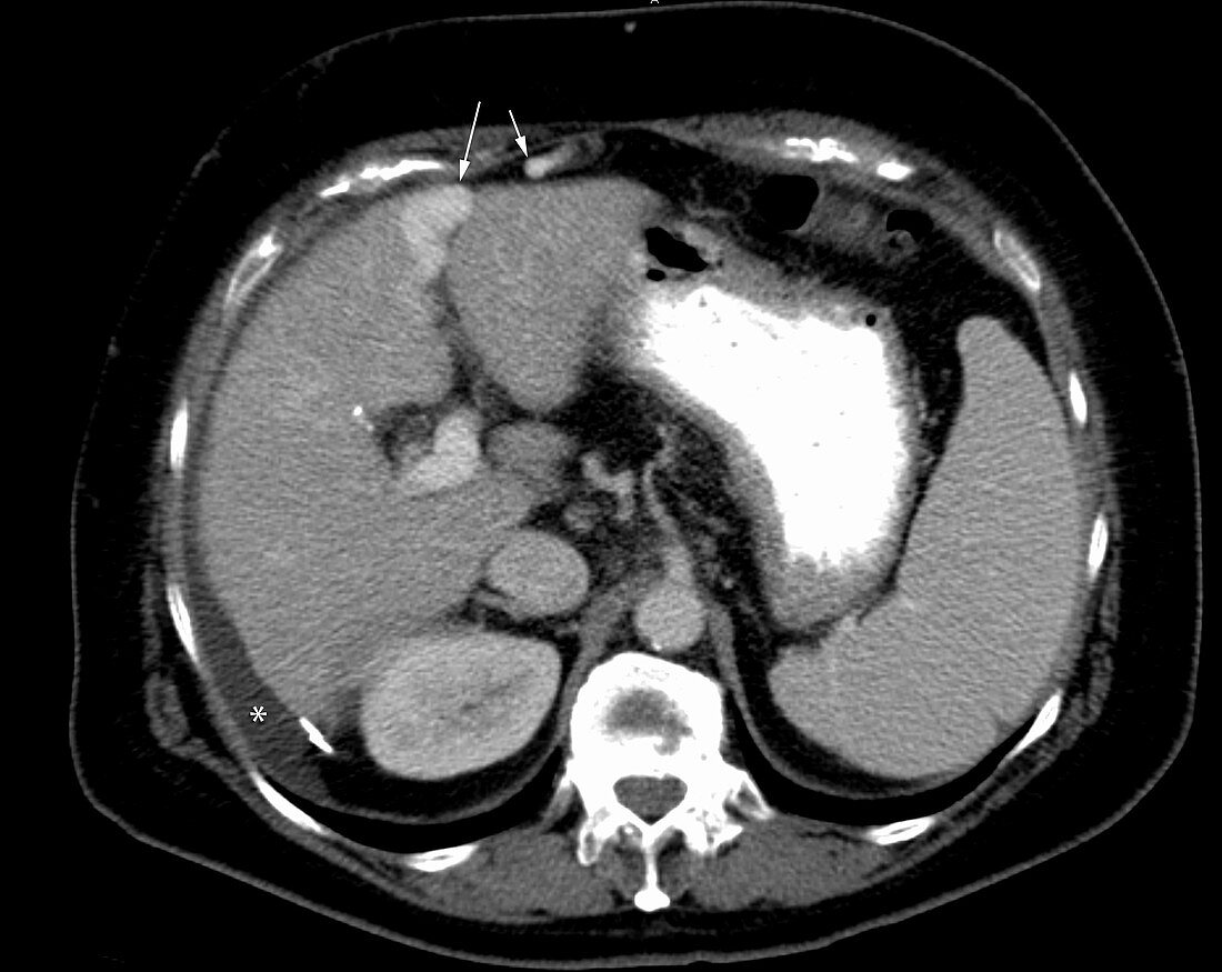
| Hepatic cirrhosis in a 67 year old female. Contrast enhanced portal venous phase axial CT scan reveals a small, shrunken liver with mild scalloped contour and mildly increased density typical of chronic hepatocellular disease. Mild perihepatic ascites fluid is present. Evidence of portal hypertension with a recanalized periumbilical vein (arrows). Paraesophageal varices are seen. | |
| Lizenzart: | Lizenzpflichtig |
| Credit: | Science Photo Library / Steven Needell |
| Bildgröße: | 2854 px × 2270 px |
| Modell-Rechte: | nicht erforderlich |
| Eigentums-Rechte: | nicht erforderlich |
| Restrictions: | - |
Preise für dieses Bild ab 15 €
Universitäten & Organisationen
(Informationsmaterial Digital, Informationsmaterial Print, Lehrmaterial Digital etc.)
ab 15 €
Redaktionell
(Bücher, Bücher: Sach- und Fachliteratur, Digitale Medien (redaktionell) etc.)
ab 30 €
Werbung
(Anzeigen, Aussenwerbung, Digitale Medien, Fernsehwerbung, Karten, Werbemittel, Zeitschriften etc.)
ab 55 €
Handelsprodukte
(bedruckte Textilie, Kalender, Postkarte, Grußkarte, Verpackung etc.)
ab 75 €
Pauschalpreise
Rechtepakete für die unbeschränkte Bildnutzung in Print oder Online
ab 495 €
Keywords
- abnormal,
- Alkohol,
- Bildgebung,
- chronisch,
- ct,
- Diagnose,
- hepatisch,
- Hepatitis,
- Infektion,
- Katze,
- klein,
- Kondition,
- Kontrast,
- Krankheit,
- Leber,
- Medizin,
- medizinisch,
- Milz,
- Missbrauch,
- Narbe,
- paraösophageal,
- Pathologie,
- pathologisch,
- Phase,
- Portal,
- Radiologie,
- Scan,
- Störung,
- ungesund,
- Vene,
- venös,
- verbessert,
- vergrößert,
- viral,
- Weiblich,
- Zirrhose