Goiter, illustration
Bildnummer 12637782
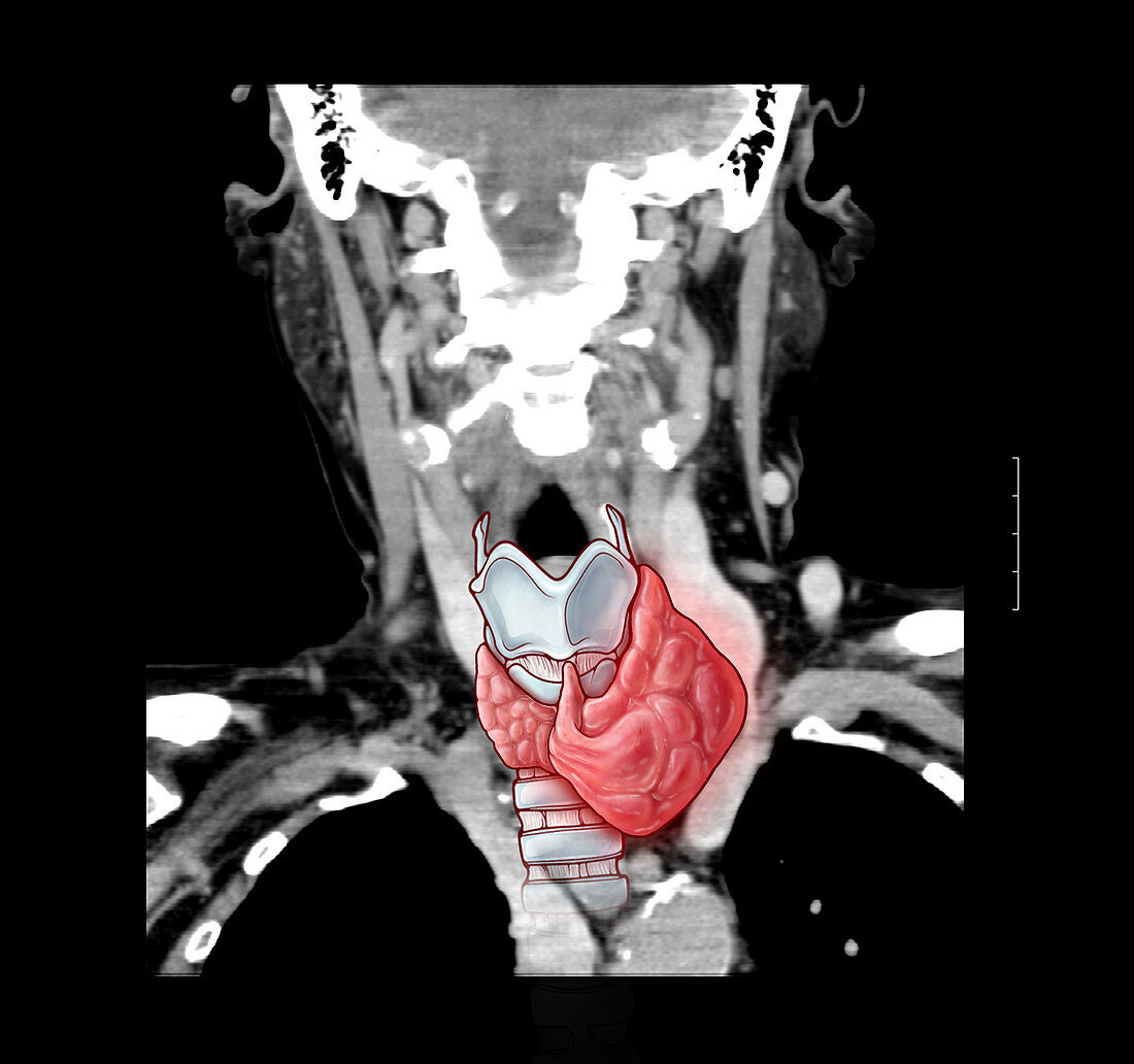
| This composit illustration of a coronal (frontal) CT reconstruction of the neck shows an abnormal left lobe of the thyroid gland which represents a thyroid goiter. This is often seen in association with iodine deficient diets. The left lobe of the thyroid is abnormally enlarged and demonstrates heterogeneous density. Also seen are the surrounding vascular structures of the superior mediastinum (central part of upper chest) and the neck. The upper lobes of the lungs are black and are on both sides. | |
| Lizenzart: | Lizenzpflichtig |
| Credit: | Science Photo Library / Oto, Evan |
| Bildgröße: | 3905 px × 3665 px |
| Modell-Rechte: | nicht erforderlich |
| Eigentums-Rechte: | nicht erforderlich |
| Restrictions: | - |
Preise für dieses Bild ab 15 €
Universitäten & Organisationen
(Informationsmaterial Digital, Informationsmaterial Print, Lehrmaterial Digital etc.)
ab 15 €
Redaktionell
(Bücher, Bücher: Sach- und Fachliteratur, Digitale Medien (redaktionell) etc.)
ab 30 €
Werbung
(Anzeigen, Aussenwerbung, Digitale Medien, Fernsehwerbung, Karten, Werbemittel, Zeitschriften etc.)
ab 55 €
Handelsprodukte
(bedruckte Textilie, Kalender, Postkarte, Grußkarte, Verpackung etc.)
ab 75 €
Pauschalpreise
Rechtepakete für die unbeschränkte Bildnutzung in Print oder Online
ab 495 €