Motor Task (feet and toes), fMRI
Bildnummer 12637612
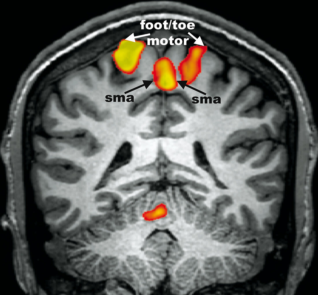
| Coronal (frontal) brain fMRI obtained while the subject was engaged in a motor task involving feet and toes. The image demonstrates normal BOLD (blood oxygen level dependent) activation (orange blobs) along the superior medial aspects of both cerebral hemispheres involving the pre and post central gyri, the motor SMA regions and minimally along the superior cerebellum. Note the more superior and medial position of brain activation compared with that generated from finger and lip motor paradigms. This is consistent with our understanding of the motor homunculus. sma = supplementary motor area | |
| Lizenzart: | Lizenzpflichtig |
| Credit: | Science Photo Library / Living Art Enterprises |
| Bildgröße: | 1618 px × 1500 px |
| Modell-Rechte: | nicht erforderlich |
| Eigentums-Rechte: | nicht erforderlich |
| Restrictions: | - |
Preise für dieses Bild ab 15 €
Universitäten & Organisationen
(Informationsmaterial Digital, Informationsmaterial Print, Lehrmaterial Digital etc.)
ab 15 €
Redaktionell
(Bücher, Bücher: Sach- und Fachliteratur, Digitale Medien (redaktionell) etc.)
ab 30 €
Werbung
(Anzeigen, Aussenwerbung, Digitale Medien, Fernsehwerbung, Karten, Werbemittel, Zeitschriften etc.)
ab 55 €
Handelsprodukte
(bedruckte Textilie, Kalender, Postkarte, Grußkarte, Verpackung etc.)
ab 75 €
Pauschalpreise
Rechtepakete für die unbeschränkte Bildnutzung in Print oder Online
ab 495 €