Finger Tapping, fMRI
Bildnummer 12637595
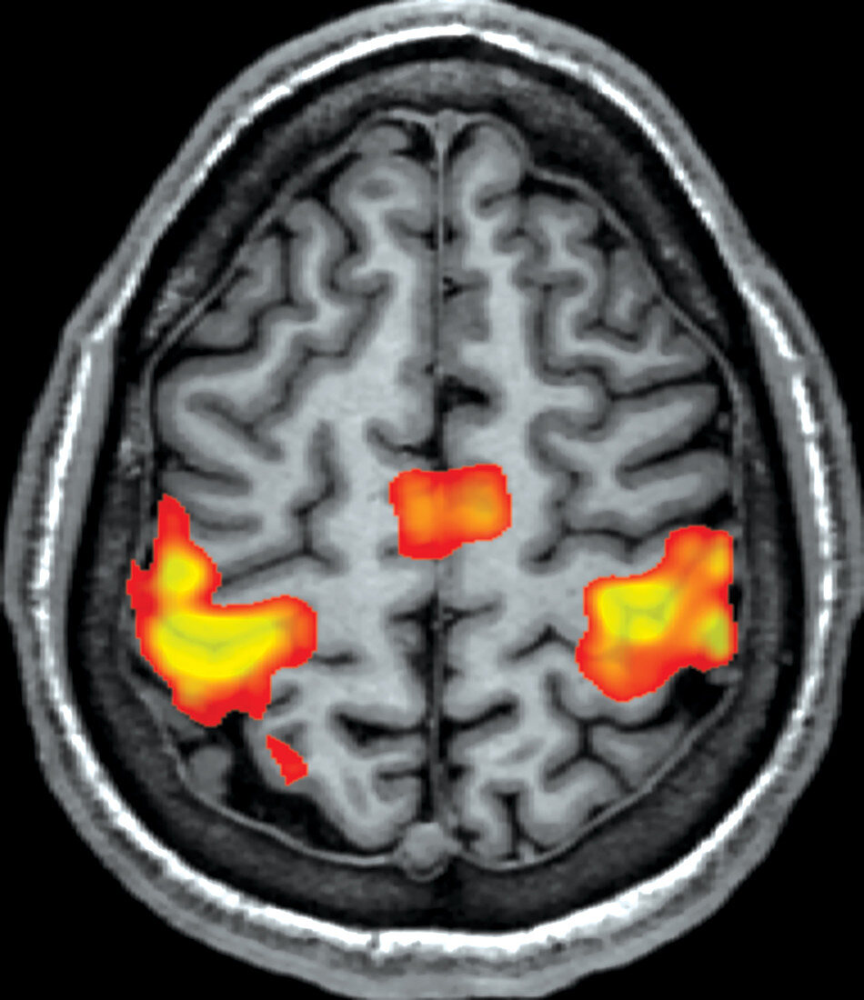
| Axial (cross sectional) fMRI of the brain during bilateral finger-tapping. Areas of BOLD (blood oxygen level dependent) activation have been overlaid on a structural 3D volume T1 weighted sequence. Normally expected brain activation is seen bilaterally in the hand motor knob regions of the pre-central gyri (primary motor area), which straddles the central sulcus extending into the post-central gyri. BOLD activation in both supplementary motor regions is seen. | |
| Lizenzart: | Lizenzpflichtig |
| Credit: | Science Photo Library / Living Art Enterprises |
| Bildgröße: | 3900 px × 4516 px |
| Modell-Rechte: | nicht erforderlich |
| Eigentums-Rechte: | nicht erforderlich |
| Restrictions: | - |
Preise für dieses Bild ab 15 €
Universitäten & Organisationen
(Informationsmaterial Digital, Informationsmaterial Print, Lehrmaterial Digital etc.)
ab 15 €
Redaktionell
(Bücher, Bücher: Sach- und Fachliteratur, Digitale Medien (redaktionell) etc.)
ab 30 €
Werbung
(Anzeigen, Aussenwerbung, Digitale Medien, Fernsehwerbung, Karten, Werbemittel, Zeitschriften etc.)
ab 55 €
Handelsprodukte
(bedruckte Textilie, Kalender, Postkarte, Grußkarte, Verpackung etc.)
ab 75 €
Pauschalpreise
Rechtepakete für die unbeschränkte Bildnutzung in Print oder Online
ab 495 €