Functional MRI, listening to music
Bildnummer 12636547
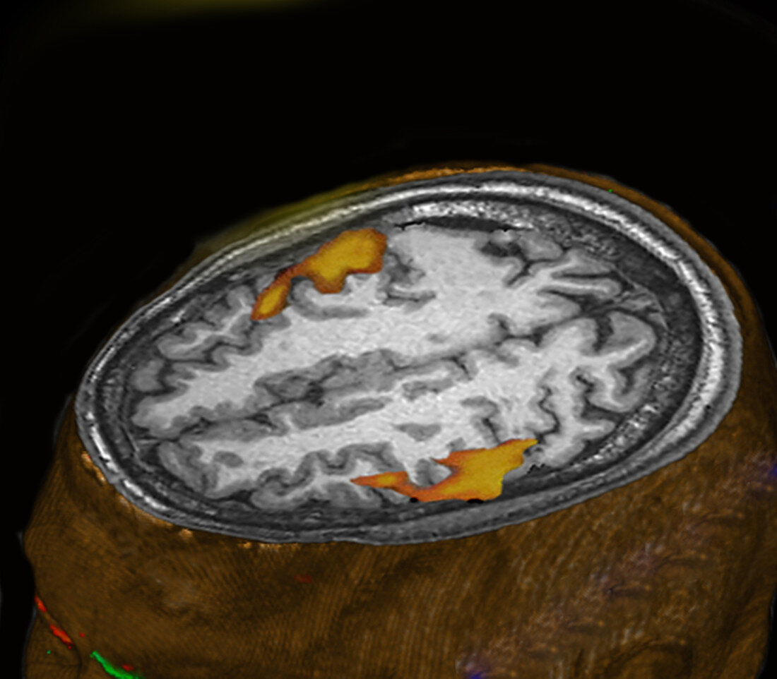
| The subject (lying in the MRI scanner) listens to instrumental music. Image shows brain areas (mostly the auditory cortex) responding to a meaningful auditory stimulus (beyond the sounds of the MRI apparatus). The small, lighter coloured areas are selected based on p=0.05 and the wider areas (orange border) represent p = 0.1. Slight blur is due to the delay in the increased flow of blood to the stimulated areas. | |
| Lizenzart: | Lizenzpflichtig |
| Credit: | Science Photo Library / Bhatia, Kul |
| Bildgröße: | 3087 px × 2700 px |
| Modell-Rechte: | nicht erforderlich |
| Eigentums-Rechte: | nicht erforderlich |
| Restrictions: | - |
Preise für dieses Bild ab 15 €
Universitäten & Organisationen
(Informationsmaterial Digital, Informationsmaterial Print, Lehrmaterial Digital etc.)
ab 15 €
Redaktionell
(Bücher, Bücher: Sach- und Fachliteratur, Digitale Medien (redaktionell) etc.)
ab 30 €
Werbung
(Anzeigen, Aussenwerbung, Digitale Medien, Fernsehwerbung, Karten, Werbemittel, Zeitschriften etc.)
ab 55 €
Handelsprodukte
(bedruckte Textilie, Kalender, Postkarte, Grußkarte, Verpackung etc.)
ab 75 €
Pauschalpreise
Rechtepakete für die unbeschränkte Bildnutzung in Print oder Online
ab 495 €