Submandibular Duct Stone, CT scan
Bildnummer 12628784
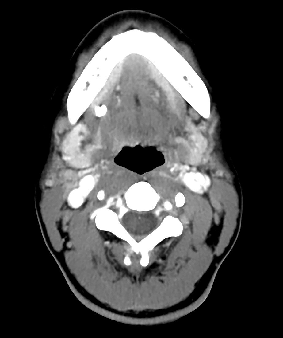
| This axial (cross sectional) contrast enhanced CT image of the head and neck demonstrates dilatation of ducts within the submandibular gland secondary to an obstructing stone (calculus) in the floor of the mouth within the submandibular duct (Wharton's duct). | |
| Lizenzart: | Lizenzpflichtig |
| Credit: | Science Photo Library / Living Art Enterprises |
| Bildgröße: | 3600 px × 4291 px |
| Modell-Rechte: | nicht erforderlich |
| Eigentums-Rechte: | nicht erforderlich |
| Restrictions: | - |
Preise für dieses Bild ab 15 €
Universitäten & Organisationen
(Informationsmaterial Digital, Informationsmaterial Print, Lehrmaterial Digital etc.)
ab 15 €
Redaktionell
(Bücher, Bücher: Sach- und Fachliteratur, Digitale Medien (redaktionell) etc.)
ab 30 €
Werbung
(Anzeigen, Aussenwerbung, Digitale Medien, Fernsehwerbung, Karten, Werbemittel, Zeitschriften etc.)
ab 55 €
Handelsprodukte
(bedruckte Textilie, Kalender, Postkarte, Grußkarte, Verpackung etc.)
ab 75 €
Pauschalpreise
Rechtepakete für die unbeschränkte Bildnutzung in Print oder Online
ab 495 €