Langerhans Cell Histiocytosis, MRI
Bildnummer 12628776
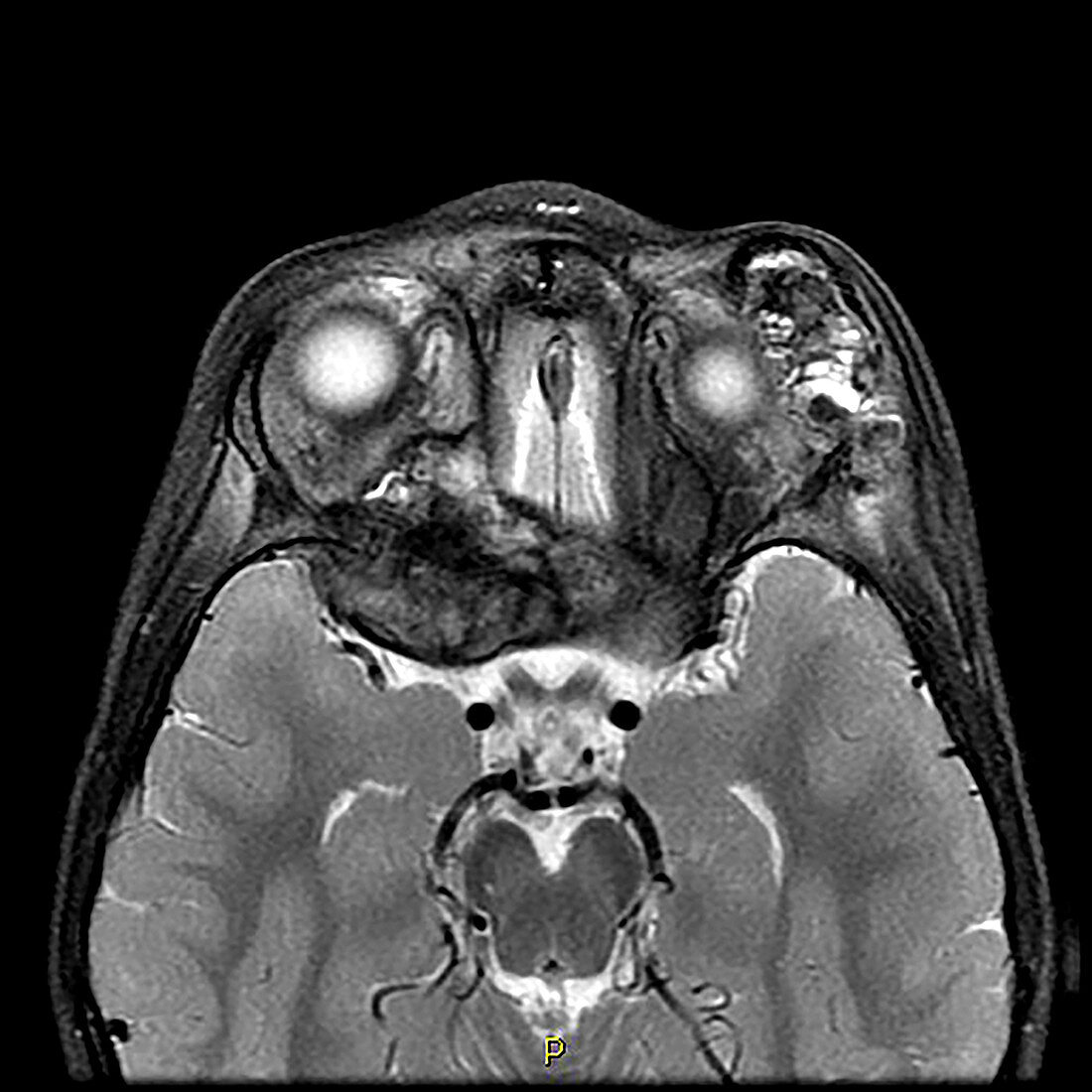
| This axial (cross sectional) T2 weighted MR image shows a destructive mass involving the right (on viewer's left) sphenoid bone, posterior orbit and region of the superior orbital fissure, representing Langerhans cell histiocytosis. There is also a lesion in the supero-lateral aspect of the left (on viewer's right) orbit. These lesions demonstrate both hyper and hypointense signal on this T2 weighted image. This disorder represents the clonal proliferation of specific cells which can be isolated to bone, or be multisystem. This disease typically affects children and adolescents. | |
| Lizenzart: | Lizenzpflichtig |
| Credit: | Science Photo Library / Living Art Enterprises |
| Bildgröße: | 3600 px × 3600 px |
| Modell-Rechte: | nicht erforderlich |
| Eigentums-Rechte: | nicht erforderlich |
| Restrictions: | - |
Preise für dieses Bild ab 15 €
Universitäten & Organisationen
(Informationsmaterial Digital, Informationsmaterial Print, Lehrmaterial Digital etc.)
ab 15 €
Redaktionell
(Bücher, Bücher: Sach- und Fachliteratur, Digitale Medien (redaktionell) etc.)
ab 30 €
Werbung
(Anzeigen, Aussenwerbung, Digitale Medien, Fernsehwerbung, Karten, Werbemittel, Zeitschriften etc.)
ab 55 €
Handelsprodukte
(bedruckte Textilie, Kalender, Postkarte, Grußkarte, Verpackung etc.)
ab 75 €
Pauschalpreise
Rechtepakete für die unbeschränkte Bildnutzung in Print oder Online
ab 495 €