Chronic Post-Traumatic Brain Injury, MRI
Bildnummer 12628453
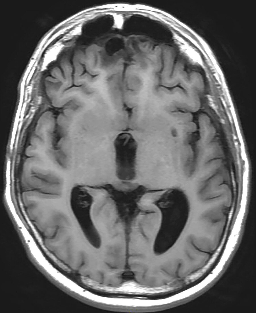
| This axial (cross sectional) T1 weighted MRI image demonstrates areas of chronic post-traumatic encephalomalacia (brain damage) in a typical location in the frontal lobes (indicated by the red arrows). Because the frontal lobes are so close to the adjacent bony skull base the brain hits the bone during the injury and then over time becomes atrophic and gliotic (brains response to injury). In the chronic phase the area of injury on MRI shows lower (looks darker) signal on T1 (as in this image) and higher (brighter or whiter) signal on T2 weighted images. | |
| Lizenzart: | Lizenzpflichtig |
| Credit: | Science Photo Library / Living Art Enterprises |
| Bildgröße: | 3600 px × 4396 px |
| Modell-Rechte: | nicht erforderlich |
| Eigentums-Rechte: | nicht erforderlich |
| Restrictions: | - |
Preise für dieses Bild ab 15 €
Universitäten & Organisationen
(Informationsmaterial Digital, Informationsmaterial Print, Lehrmaterial Digital etc.)
ab 15 €
Redaktionell
(Bücher, Bücher: Sach- und Fachliteratur, Digitale Medien (redaktionell) etc.)
ab 30 €
Werbung
(Anzeigen, Aussenwerbung, Digitale Medien, Fernsehwerbung, Karten, Werbemittel, Zeitschriften etc.)
ab 55 €
Handelsprodukte
(bedruckte Textilie, Kalender, Postkarte, Grußkarte, Verpackung etc.)
ab 75 €
Pauschalpreise
Rechtepakete für die unbeschränkte Bildnutzung in Print oder Online
ab 495 €