Pancreatic Islet Stained for A Cells, LM
Bildnummer 12627520
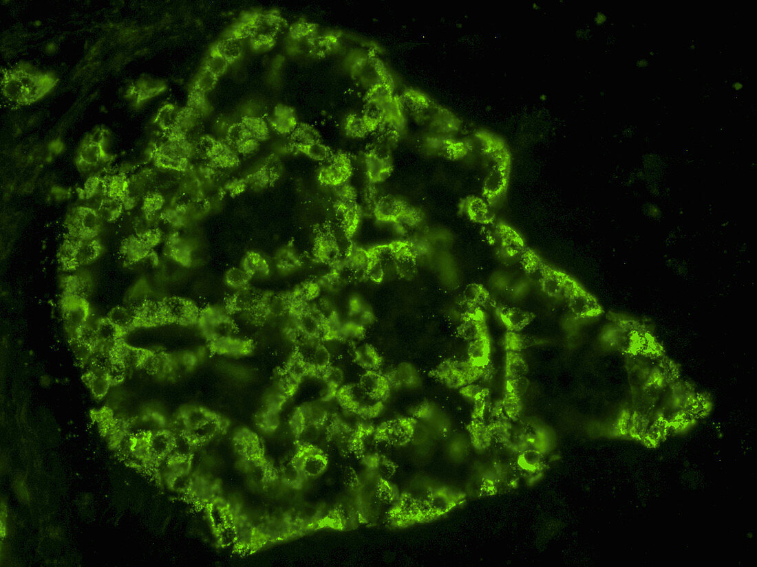
| Light micrograph of a pancreatic islet stained with an antibody to the major peptide hormone glucagon to show the relative content and distribution of the alpha, or A cells, which produce it. The antibody was labelled with a green fluorescent dye. One will note that there are a few fluorescent cells, outside the islet, that stain one of the three colours. These are either isolated islet cells that 1) are not confined to the islet, 2) belong to an islet almost entirely outside the plane of the section, or 3) are an artefact of sectioning, which was done on frozen tissue. Magnification: 400X. | |
| Lizenzart: | Lizenzpflichtig |
| Credit: | Science Photo Library / Alvin Telser |
| Bildgröße: | 4000 px × 3000 px |
| Modell-Rechte: | nicht erforderlich |
| Eigentums-Rechte: | nicht erforderlich |
| Restrictions: | - |
Preise für dieses Bild ab 15 €
Universitäten & Organisationen
(Informationsmaterial Digital, Informationsmaterial Print, Lehrmaterial Digital etc.)
ab 15 €
Redaktionell
(Bücher, Bücher: Sach- und Fachliteratur, Digitale Medien (redaktionell) etc.)
ab 30 €
Werbung
(Anzeigen, Aussenwerbung, Digitale Medien, Fernsehwerbung, Karten, Werbemittel, Zeitschriften etc.)
ab 55 €
Handelsprodukte
(bedruckte Textilie, Kalender, Postkarte, Grußkarte, Verpackung etc.)
ab 75 €
Pauschalpreise
Rechtepakete für die unbeschränkte Bildnutzung in Print oder Online
ab 495 €