Enterobacteria phage T2
Bildnummer 12627236
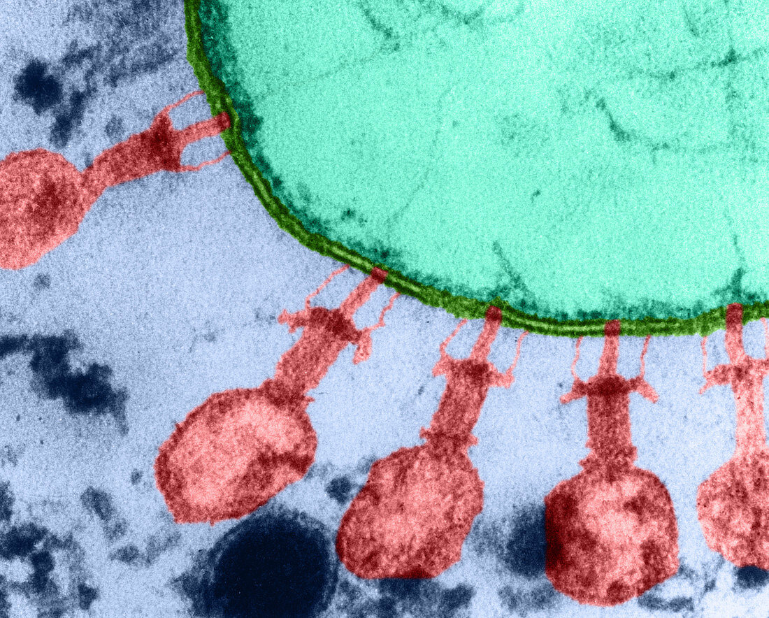
| Colour enhanced transmission electron micrograph showing sections through absorbed T2 phages attached to intact E. coli B cells or to large wall fragments by both their long and short tail fibres. E. coli B cells were infected with T2 phages; after 1 hour they were fixed and then embedded. Rather thick, silvery gold-coloured sections were cut and subsequently examined in the electron microscope. The long tail fibres extend laterally from the baseplates; they flex near their centers and their distal tips are attached to the cell walls. The long tail fibres attach to the walls at points over 800 A from the intersections of the needles with the walls. The short tail fibres go directly from the baseplates to the walls. | |
| Lizenzart: | Lizenzpflichtig |
| Credit: | Science Photo Library / Simon, Lee D. |
| Bildgröße: | 3677 px × 2956 px |
| Modell-Rechte: | nicht erforderlich |
| Eigentums-Rechte: | nicht erforderlich |
| Restrictions: | - |
Preise für dieses Bild ab 15 €
Universitäten & Organisationen
(Informationsmaterial Digital, Informationsmaterial Print, Lehrmaterial Digital etc.)
ab 15 €
Redaktionell
(Bücher, Bücher: Sach- und Fachliteratur, Digitale Medien (redaktionell) etc.)
ab 30 €
Werbung
(Anzeigen, Aussenwerbung, Digitale Medien, Fernsehwerbung, Karten, Werbemittel, Zeitschriften etc.)
ab 55 €
Handelsprodukte
(bedruckte Textilie, Kalender, Postkarte, Grußkarte, Verpackung etc.)
ab 75 €
Pauschalpreise
Rechtepakete für die unbeschränkte Bildnutzung in Print oder Online
ab 495 €
Keywords
- Bakterien,
- Bakteriophage,
- Bakterium,
- E coli,
- E. coli,
- eingefärbt,
- elektronenmikroskopische Aufnahme,
- Escherichia coli,
- Gastgeber,
- medizinisch,
- Mikrobiologie,
- Mikrofotografie,
- Mikroskopie,
- Mikroskopische Aufnahmen,
- t2,
- tem,
- transmissionselektronenmikroskopische Aufnahme,
- verbessert,
- viral,
- Virus,
- Wissenschaft