Mitochondrion in intestinal cell, FIB-SEM image
Bildnummer 12581320
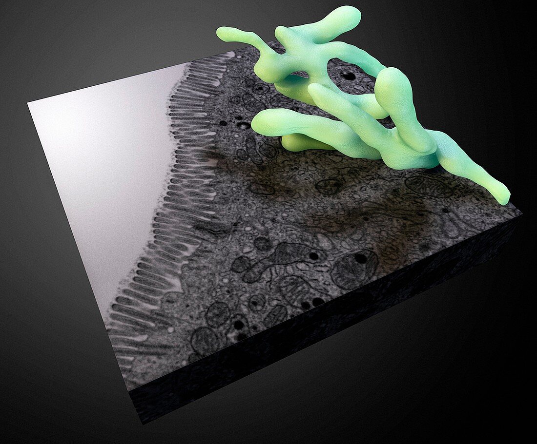
| Mitochondrion in intestinal cell, FIB-SEM image. Three-dimensional structure of a single mitochondrion in a cell lining the small intestine, shown with the stack of electron micrographs that it was derived from. This image was produced using a technique called focused ion beam scanning electron microscopy (FIB-SEM).Image created in September 2014. | |
| Lizenzart: | Lizenzpflichtig |
| Credit: | Science Photo Library / National Cancer Institute / Amy Moran (Nlm), Sriram Subramaniam |
| Bildgröße: | 3253 px × 2694 px |
| Modell-Rechte: | nicht erforderlich |
| Eigentums-Rechte: | nicht erforderlich |
| Restrictions: | - |
Preise für dieses Bild ab 15 €
Universitäten & Organisationen
(Informationsmaterial Digital, Informationsmaterial Print, Lehrmaterial Digital etc.)
ab 15 €
Redaktionell
(Bücher, Bücher: Sach- und Fachliteratur, Digitale Medien (redaktionell) etc.)
ab 30 €
Werbung
(Anzeigen, Aussenwerbung, Digitale Medien, Fernsehwerbung, Karten, Werbemittel, Zeitschriften etc.)
ab 55 €
Handelsprodukte
(bedruckte Textilie, Kalender, Postkarte, Grußkarte, Verpackung etc.)
ab 75 €
Pauschalpreise
Rechtepakete für die unbeschränkte Bildnutzung in Print oder Online
ab 495 €
Keywords
- 2014,
- 21. Jahrhundert,
- 3 dimensional,
- 3-d,
- 3-dimensional,
- 3D,
- ausgeschnitten,
- Ausschnitte,
- Biologie,
- biologisch,
- Dreidimensional,
- Dünndarm,
- farbig,
- gefärbt,
- menschlicher Körper,
- Mitochondrion,
- Niemand,
- Organelle,
- Rasterelektronenmikroskopie,
- rasterelektronenmikroskopische Aufnahme,
- REM,
- schwarzer Hintergrund,
- Zellbilogie,
- Zelle,
- zellular,
- Zytologie,
- Zytologisch