Opioid receptor distribution in rat brain, light micrograph
Bildnummer 12540076
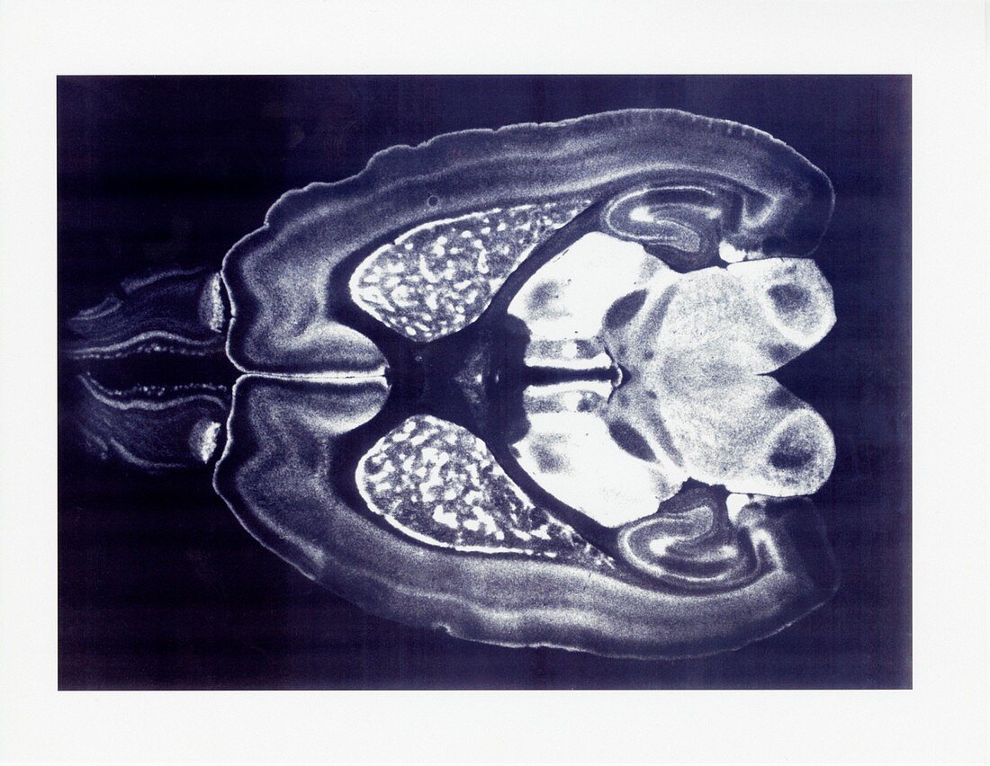
| Opioid receptor distribution, light micrograph. Autoradiographic visualization with dark-field illumination of mu opioid receptor distribution in a horizontal brain section from a rat. White areas show intricate patterns of opioid receptor distribution in well-defined brain areas. These include the olfactory bulb, the cerebral cortex, the hippocampus, the striatum, the thalamus, and the tectum. Receptors are marked by deuterated naloxone binding, followed by autoradiographic visualization of emulsion-coated brain sections. | |
| Lizenzart: | Lizenzpflichtig |
| Credit: | Science Photo Library / Miles Herkenham, NIMH / National Institute on Drug Abuse, NATIONAL INSTITUTES OF HEALTH |
| Bildgröße: | 3360 px × 2601 px |
| Modell-Rechte: | nicht erforderlich |
| Eigentums-Rechte: | nicht erforderlich |
| Restrictions: | - |
Preise für dieses Bild ab 15 €
Universitäten & Organisationen
(Informationsmaterial Digital, Informationsmaterial Print, Lehrmaterial Digital etc.)
ab 15 €
Redaktionell
(Bücher, Bücher: Sach- und Fachliteratur, Digitale Medien (redaktionell) etc.)
ab 30 €
Werbung
(Anzeigen, Aussenwerbung, Digitale Medien, Fernsehwerbung, Karten, Werbemittel, Zeitschriften etc.)
ab 55 €
Handelsprodukte
(bedruckte Textilie, Kalender, Postkarte, Grußkarte, Verpackung etc.)
ab 75 €
Pauschalpreise
Rechtepakete für die unbeschränkte Bildnutzung in Print oder Online
ab 495 €
Keywords
- Autoradiogramm,
- Autoradiographie,
- Biologie,
- biologisch,
- Einfarbig,
- Gehirn,
- gesund,
- Hirnrinde,
- Lichtmikroskop,
- lichtmikroskopische Aufnahme,
- Natur,
- neural,
- Neurologie,
- neurologisch,
- Niemand,
- normal,
- Opioidrezeptoren,
- Organ,
- Physiologie,
- physiologisch,
- Querschnitt,
- Ratte,
- Riechkolben,
- Schwarz und weiß,
- Sektion,
- sektioniert,
- Thalamus,
- Tier,
- Zoologie,
- zoologisch