Nasal epithelium, SEM
Bildnummer 12540044
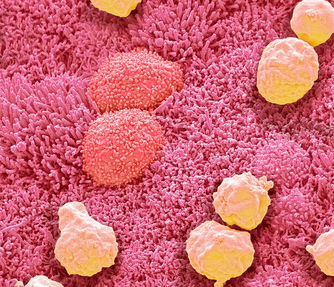
| Nasal epithelium. Coloured scanning electron micrograph (SEM). Shown here are squamous nasal epithelial cells (red) and mucous (yellow). The epithelial cell surfaces are covered with tiny microvilli that increase the cell surface area. The microvilli likely aid in localization of foreign debris (coming from the nose) and detection by immune cells. Mucus, secreted by cells in the epithelial lining traps foreign objects, such as bacteria, preventing them from entering the lungs. Magnification: x4000 when printed at 10 centimetres wide. | |
| Lizenzart: | Lizenzpflichtig |
| Credit: | Science Photo Library / Gschmeissner, Steve |
| Bildgröße: | 4572 px × 3938 px |
| Modell-Rechte: | nicht erforderlich |
| Eigentums-Rechte: | nicht erforderlich |
| Restrictions: | - |
Preise für dieses Bild ab 15 €
Universitäten & Organisationen
(Informationsmaterial Digital, Informationsmaterial Print, Lehrmaterial Digital etc.)
ab 15 €
Redaktionell
(Bücher, Bücher: Sach- und Fachliteratur, Digitale Medien (redaktionell) etc.)
ab 30 €
Werbung
(Anzeigen, Aussenwerbung, Digitale Medien, Fernsehwerbung, Karten, Werbemittel, Zeitschriften etc.)
ab 55 €
Handelsprodukte
(bedruckte Textilie, Kalender, Postkarte, Grußkarte, Verpackung etc.)
ab 75 €
Pauschalpreise
Rechtepakete für die unbeschränkte Bildnutzung in Print oder Online
ab 495 €