Small intestine villi, SEM
Bildnummer 12528279
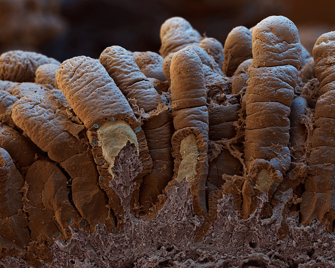
| Small intestine villi. Coloured scanning electron micrograph (SEM) of villi (brown) on the lining of the small intestine. Villi greatly increase the surface area for the absorption of nutrients from food. Several of the villi have been partially broken, revealing their internal structure. The villus epithelium (outer layer) contains enterocytes (brown, columnar cells), which are involved in nutrient absorption. Scattered amongst these are goblet cells (blue), which secrete mucus onto the intestinal surface. The inner villus tissue (yellow/grey) contains blood vessels which transport digestive products to a nearby vein. Magnification: x62 at 6x7cm size. x100 at 4x5ins | |
| Lizenzart: | Lizenzpflichtig |
| Credit: | Science Photo Library / EYE OF SCIENCE |
| Bildgröße: | 4000 px × 3200 px |
| Modell-Rechte: | nicht erforderlich |
| Eigentums-Rechte: | nicht erforderlich |
| Restrictions: |
|
Preise für dieses Bild ab 15 €
Universitäten & Organisationen
(Informationsmaterial Digital, Informationsmaterial Print, Lehrmaterial Digital etc.)
ab 15 €
Redaktionell
(Bücher, Bücher: Sach- und Fachliteratur, Digitale Medien (redaktionell) etc.)
ab 30 €
Werbung
(Anzeigen, Aussenwerbung, Digitale Medien, Fernsehwerbung, Karten, Werbemittel, Zeitschriften etc.)
ab 55 €
Handelsprodukte
(bedruckte Textilie, Kalender, Postkarte, Grußkarte, Verpackung etc.)
ab 75 €
Pauschalpreise
Rechtepakete für die unbeschränkte Bildnutzung in Print oder Online
ab 495 €
Keywords
- Absorption,
- Anatomie,
- Darm,
- Dünndarm,
- Enterozyten,
- farbig,
- Futter,
- gesund,
- Gewebe,
- kaputt,
- menschlicher Körper,
- Nährstoff,
- normal,
- Projektion,
- Projektionen,
- rasterelektronenmikroskopische Aufnahme,
- REM,
- Schleimhaut,
- sekretorisch,
- Sektion,
- sektioniert,
- Standort,
- Trakt,
- vaskulär,
- Verdauung,
- Verdauungskanal,
- Verdauungssystem,
- Zellen,
- Zotte,
- Zotten