Slime mould fruiting bodies, SEM
Bildnummer 12525660
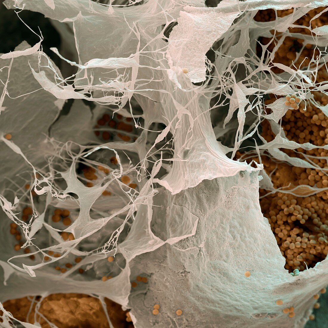
| Slime mould fruiting body. Coloured scanning electron micrograph (SEM) of the fruiting body (sporangium) of the slime mould Badhamia utricularis. The delicate outer wall (peridium, white) of the sporangium has burst, releasing spores (small orange spheres). The spores are associated with a mass of threads (the capillitium, thin white strands), which in this species contain calcium oxide. Changes in the moisture content of the air cause these threads to change shape, flicking spores into the air and helping them disperse. Slime moulds are not fungi, but a separate group with complex life cycles. Magnification: x150 at 6x6cm size. | |
| Lizenzart: | Lizenzpflichtig |
| Credit: | Science Photo Library / EYE OF SCIENCE |
| Bildgröße: | 4000 px × 4000 px |
| Modell-Rechte: | nicht erforderlich |
| Eigentums-Rechte: | nicht erforderlich |
| Restrictions: |
|
Preise für dieses Bild ab 15 €
Universitäten & Organisationen
(Informationsmaterial Digital, Informationsmaterial Print, Lehrmaterial Digital etc.)
ab 15 €
Redaktionell
(Bücher, Bücher: Sach- und Fachliteratur, Digitale Medien (redaktionell) etc.)
ab 30 €
Werbung
(Anzeigen, Aussenwerbung, Digitale Medien, Fernsehwerbung, Karten, Werbemittel, Zeitschriften etc.)
ab 55 €
Handelsprodukte
(bedruckte Textilie, Kalender, Postkarte, Grußkarte, Verpackung etc.)
ab 75 €
Pauschalpreise
Rechtepakete für die unbeschränkte Bildnutzung in Print oder Online
ab 495 €