Coloured PET brain scan during olfactory activity
Bildnummer 12496935
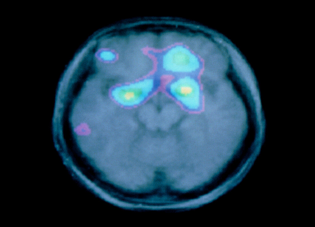
| Brain's olfactory centre. Coloured positron emission tomography (PET) scan of the brain during olfactory (smell) activity. The PET scan has been superimposed onto a grey magnetic resonance imaging (MRI) scan. In this axial (horizontal) section the front of the brain is at top. The scan shows the level of brain activity associated with olfactory activity from low (purple) to high (yellow). The highest activity is in the olfactory centres (green & yellow) of the sensory cortex. PET scans use radioactively- labelled substances introduced into the blood to view metabolic activity. | |
| Lizenzart: | Lizenzpflichtig |
| Credit: | Science Photo Library / MONTREAL NEUROLOGICAL INSTITUTE |
| Bildgröße: | 3543 px × 2551 px |
| Modell-Rechte: | nicht erforderlich |
| Eigentums-Rechte: | nicht erforderlich |
| Restrictions: | - |
Preise für dieses Bild ab 15 €
Universitäten & Organisationen
(Informationsmaterial Digital, Informationsmaterial Print, Lehrmaterial Digital etc.)
ab 15 €
Redaktionell
(Bücher, Bücher: Sach- und Fachliteratur, Digitale Medien (redaktionell) etc.)
ab 30 €
Werbung
(Anzeigen, Aussenwerbung, Digitale Medien, Fernsehwerbung, Karten, Werbemittel, Zeitschriften etc.)
ab 55 €
Handelsprodukte
(bedruckte Textilie, Kalender, Postkarte, Grußkarte, Verpackung etc.)
ab 75 €
Pauschalpreise
Rechtepakete für die unbeschränkte Bildnutzung in Print oder Online
ab 495 €