Coloured 3-D PET brain scan during visual activity
Bildnummer 12496929
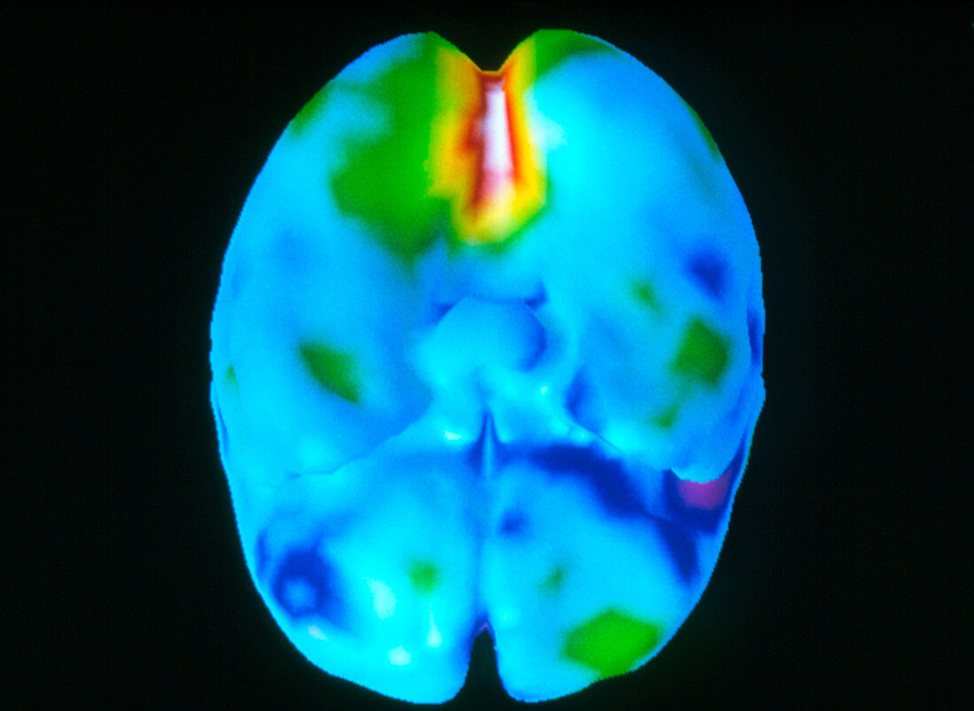
| Brain's visual centre. Coloured three-dimensional positron emission tomography (PET) scan of the brain seen from below during visual activity. The frontal lobe is at lower centre. The scan shows the amount of glucose (sugar) consumption and hence areas of low (blue) and high (red & white) brain activity. The most active area is the visual cortex within the occipital lobe at the back of the brain (at upper centre). The cerebellum has been removed to allow this area to be viewed more clearly. PET scans use radioactively-labelled substances introduced into the blood to view metabolic activity in three-dimensions. | |
| Lizenzart: | Lizenzpflichtig |
| Credit: | Science Photo Library / MONTREAL NEUROLOGICAL INSTITUTE |
| Bildgröße: | 3543 px × 2588 px |
| Modell-Rechte: | nicht erforderlich |
| Eigentums-Rechte: | nicht erforderlich |
| Restrictions: | - |
Preise für dieses Bild ab 15 €
Universitäten & Organisationen
(Informationsmaterial Digital, Informationsmaterial Print, Lehrmaterial Digital etc.)
ab 15 €
Redaktionell
(Bücher, Bücher: Sach- und Fachliteratur, Digitale Medien (redaktionell) etc.)
ab 30 €
Werbung
(Anzeigen, Aussenwerbung, Digitale Medien, Fernsehwerbung, Karten, Werbemittel, Zeitschriften etc.)
ab 55 €
Handelsprodukte
(bedruckte Textilie, Kalender, Postkarte, Grußkarte, Verpackung etc.)
ab 75 €
Pauschalpreise
Rechtepakete für die unbeschränkte Bildnutzung in Print oder Online
ab 495 €