Comparison of gross anatomy of acute and chronic pyelonephri
Bildnummer 12496753
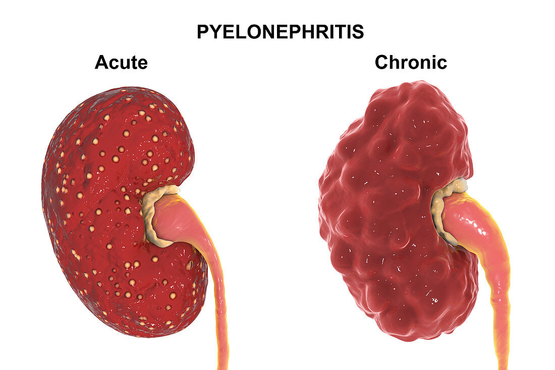
| Comparison of gross anatomy of acute and chronic pyelonephritis, illustration. There are small abscesses (yellow) on the surface of the kidney with acute pyelonephritis (left). The kidney with chronic pyelonephritis (right) has an irregular scarred cortical surface, dilated and blunted calyces (yellow), and a dilated ureter. | |
| Lizenzart: | Lizenzfrei |
| Credit: | Science Photo Library / Kon, Kateryna |
| Modell-Rechte: | nicht erforderlich |
| Eigentums-Rechte: | nicht erforderlich |
| Restrictions: | - |
Preise für dieses Bild ab 29 €
Für digitale Nutzung (72 dpi)
ab 29 €
Für Druckauflösung (300 dpi)
ab 300 €
Keywords
- 3 dimensional,
- 3D,
- abnormal,
- Abszess,
- Akut,
- anatomische Darstellung,
- bakteriell,
- Biologie,
- biologisch,
- chronisch,
- digital generiert,
- Dreidimensional,
- einer,
- einfacher Hintergrund,
- Erkrankung,
- Gesundheitswesen und Medizin,
- Harnleiter,
- Harnsystem,
- Illustration,
- Infektion,
- infiziert,
- Kondition,
- Kontraste,
- krank,
- Krankheit,
- Kunstwerk,
- Medizin,
- medizinisch,
- menschlicher Körper,
- menschliches Organ,
- Nephrologie,
- Niemand,
- Niere,
- Nieren-,
- Organ,
- Single,
- Störung,
- ungesund,
- Vergleich,
- Vernarbung,
- weißer Hintergrund