Inner ear and brain structures, 3D MRI scan
Bildnummer 12491662
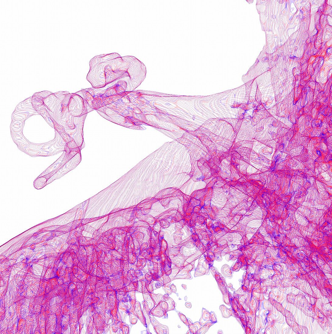
| Inner ear and brain structures. Coloured 3D magnetic resonance imaging (MRI) scan of the anatomy of the inner ear and associated brain structures. This view from above is of a normal left ear, showing the internal auditory meatus (IAM) at upper centre, with the inner ear at upper left. The cranial nerves (CN VII, facial nerve; and CN VIII, vestibulocochlear nerve) are seen crossing the cerebellopontine angle (CPA) at centre, which is between the cerebellum and the pons. The pink areas at right and bottom are parts of the brain. | |
| Lizenzart: | Lizenzpflichtig |
| Credit: | Science Photo Library / Fung, K.H. |
| Bildgröße: | 4207 px × 4237 px |
| Modell-Rechte: | nicht erforderlich |
| Eigentums-Rechte: | nicht erforderlich |
| Restrictions: | - |
Preise für dieses Bild ab 15 €
Universitäten & Organisationen
(Informationsmaterial Digital, Informationsmaterial Print, Lehrmaterial Digital etc.)
ab 15 €
Redaktionell
(Bücher, Bücher: Sach- und Fachliteratur, Digitale Medien (redaktionell) etc.)
ab 30 €
Werbung
(Anzeigen, Aussenwerbung, Digitale Medien, Fernsehwerbung, Karten, Werbemittel, Zeitschriften etc.)
ab 55 €
Handelsprodukte
(bedruckte Textilie, Kalender, Postkarte, Grußkarte, Verpackung etc.)
ab 75 €
Pauschalpreise
Rechtepakete für die unbeschränkte Bildnutzung in Print oder Online
ab 495 €
Keywords
- 3 dimensional,
- 3-d,
- 3-dimensional,
- 3D,
- Acht,
- Anatomie,
- anatomisch,
- Audiologie,
- aural,
- Balance,
- Biologie,
- biologisch,
- Dreidimensional,
- farbig,
- gefärbt,
- Gehirn,
- gesund,
- Hirnnerven,
- Hören,
- Innenohr,
- Kleinhirn,
- Magnetresonanztomografie,
- menschlicher Körper,
- MRT-Untersuchung,
- Niemand,
- normal,
- Ohr,
- Organ,
- rosa,
- Scanner,
- sensorisch,
- Sinn,
- Struktur,
- Vorhalle,
- weißer Hintergrund