Skin mole, light micrograph
Bildnummer 12421699
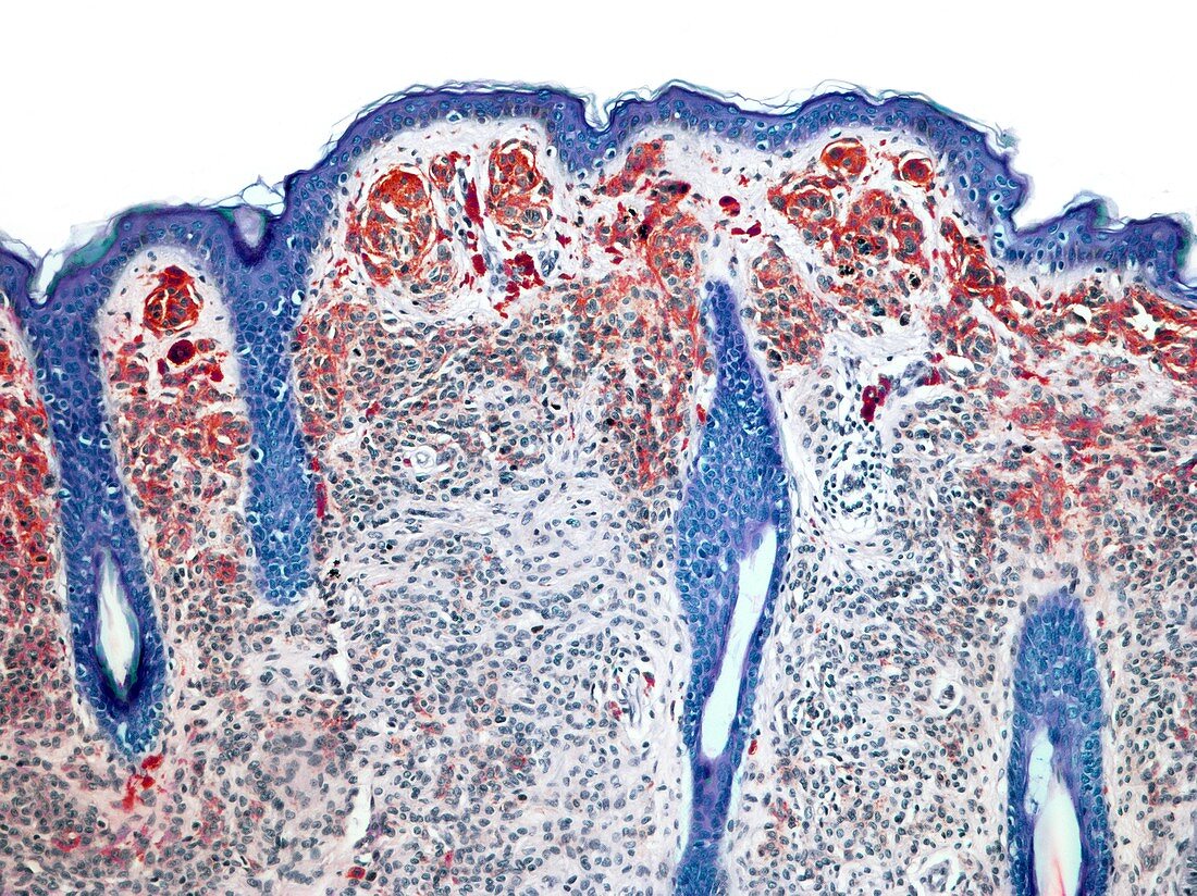
| Skin mole. Light micrograph (LM) of a section through a naevus, a type of skin mole. This pigmented naevus is produced by abnormal clustering of pigment cells (melanocytes red) in the skin. At top are loose cornified layers on the surface of the skin. The epidermis is immediately below this, followed by the dermis. Throughout this region there are clusters of red melanocyte pigment cells. Naevi are harmless, but on rare occasions can become cancerous. Magnification: x15 when printed at 10 centimetres wide. Human tissue. | |
| Lizenzart: | Lizenzpflichtig |
| Credit: | Science Photo Library / Gschmeissner, Steve |
| Bildgröße: | 4829 px × 3619 px |
| Modell-Rechte: | nicht erforderlich |
| Eigentums-Rechte: | nicht erforderlich |
| Restrictions: | - |
Preise für dieses Bild ab 15 €
Universitäten & Organisationen
(Informationsmaterial Digital, Informationsmaterial Print, Lehrmaterial Digital etc.)
ab 15 €
Redaktionell
(Bücher, Bücher: Sach- und Fachliteratur, Digitale Medien (redaktionell) etc.)
ab 30 €
Werbung
(Anzeigen, Aussenwerbung, Digitale Medien, Fernsehwerbung, Karten, Werbemittel, Zeitschriften etc.)
ab 55 €
Handelsprodukte
(bedruckte Textilie, Kalender, Postkarte, Grußkarte, Verpackung etc.)
ab 75 €
Pauschalpreise
Rechtepakete für die unbeschränkte Bildnutzung in Print oder Online
ab 495 €