Nasal lining, SEM
Bildnummer 12420790
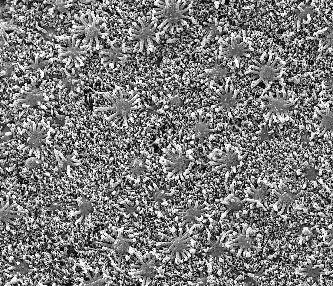
| Nasal lining. Scanning electron micrograph (SEM) of the olfactory epithelium that lines the nasal cavity, showing olfactory cells (star shaped) surrounded by numerous cilia (hair-like projections). The cilia are covered with a sticky mucus (not seen) that traps dust and other inhaled particles. Co-ordinated, wave-like beating of the cilia propels the mucus to the back of the nose (pharynx), where it is swallowed. The olfactory vesicles are bipolar neurons (nerve cells) that transmit 'smell' information to the brain. Magnification: x300 when printed 10 centimetres wide. | |
| Lizenzart: | Lizenzpflichtig |
| Credit: | Science Photo Library / Gschmeissner, Steve |
| Bildgröße: | 4571 px × 3925 px |
| Modell-Rechte: | nicht erforderlich |
| Eigentums-Rechte: | nicht erforderlich |
| Restrictions: | - |
Preise für dieses Bild ab 15 €
Universitäten & Organisationen
(Informationsmaterial Digital, Informationsmaterial Print, Lehrmaterial Digital etc.)
ab 15 €
Redaktionell
(Bücher, Bücher: Sach- und Fachliteratur, Digitale Medien (redaktionell) etc.)
ab 30 €
Werbung
(Anzeigen, Aussenwerbung, Digitale Medien, Fernsehwerbung, Karten, Werbemittel, Zeitschriften etc.)
ab 55 €
Handelsprodukte
(bedruckte Textilie, Kalender, Postkarte, Grußkarte, Verpackung etc.)
ab 75 €
Pauschalpreise
Rechtepakete für die unbeschränkte Bildnutzung in Print oder Online
ab 495 €
Keywords
- Anatomie,
- anatomisch,
- Biologie,
- biologisch,
- Einfarbig,
- Epithel,
- epithelial,
- Futter,
- Geruch,
- Geruchssinn,
- Gewebe,
- Haar,
- Haare,
- Membran,
- Nase,
- Neurologie,
- neurologisch,
- Neuron,
- Neuronen,
- olfaktorisch,
- olfaktorisches Vesikel,
- Rasterelektronenmikroskop,
- rasterelektronenmikroskopische Aufnahme,
- REM,
- Schleimhaut,
- schwarz und weiß,
- sekretorisch,
- Sinn,
- Wimpern,
- Zelle,
- Zellen