Abdomen and chest anatomy, CT scans
Bildnummer 12398552
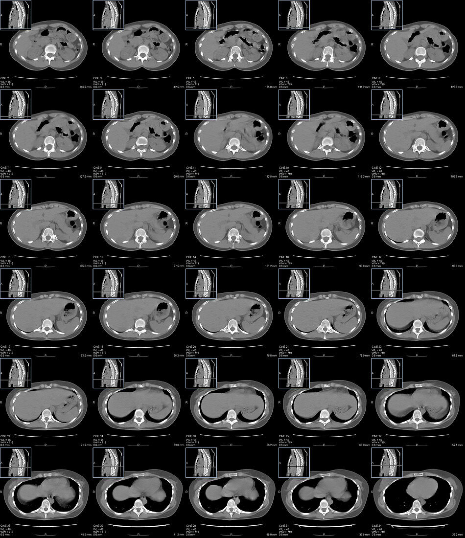
| Abdomen and chest anatomy. Series of computer tomography (CT) scans of transverse sections through the upper part of the abdomen and the lower part of chest of a 30 year old patient. They show the liver (left), bowels (right), spine (bottom), kidneys (bottom left and right), heart (top in last line of images) and lower lobes of lungs (black in last line of images). | |
| Lizenzart: | Lizenzpflichtig |
| Credit: | Science Photo Library / Kon, Kateryna |
| Bildgröße: | 3885 px × 4499 px |
| Modell-Rechte: | nicht erforderlich |
| Eigentums-Rechte: | nicht erforderlich |
| Restrictions: | - |
Preise für dieses Bild ab 15 €
Universitäten & Organisationen
(Informationsmaterial Digital, Informationsmaterial Print, Lehrmaterial Digital etc.)
ab 15 €
Redaktionell
(Bücher, Bücher: Sach- und Fachliteratur, Digitale Medien (redaktionell) etc.)
ab 30 €
Werbung
(Anzeigen, Aussenwerbung, Digitale Medien, Fernsehwerbung, Karten, Werbemittel, Zeitschriften etc.)
ab 55 €
Handelsprodukte
(bedruckte Textilie, Kalender, Postkarte, Grußkarte, Verpackung etc.)
ab 75 €
Pauschalpreise
Rechtepakete für die unbeschränkte Bildnutzung in Print oder Online
ab 495 €
Keywords
- anatomisch,
- Aorta,
- Arterie,
- Atmungssystem,
- Blutgefäß,
- Blutversorgung,
- Computertomographie,
- CT-Scan,
- Darm,
- Detail,
- direkt darüber,
- Dreidimensional,
- Dünndarm,
- Erwachsene,
- gesund,
- Gesundheitswesen,
- Herz,
- Herzkreislaufsystem,
- inneres Organ,
- Knochen,
- Leber,
- Lunge,
- Medizin,
- medizinisch,
- menschliche Anatomie,
- menschlicher Körper,
- menschliches Körperteil,
- menschliches Skelett,
- Nahansicht,
- Nieren,
- normal,
- Organ,
- Querschnitt,
- schwarzer Hintergrund,
- Serie,
- Transversalschnitt,
- Truhe,
- Wirbelsäule,
- wissenschaftliche Bildgebung