Stroke, MRI brain scans
Bildnummer 12378960
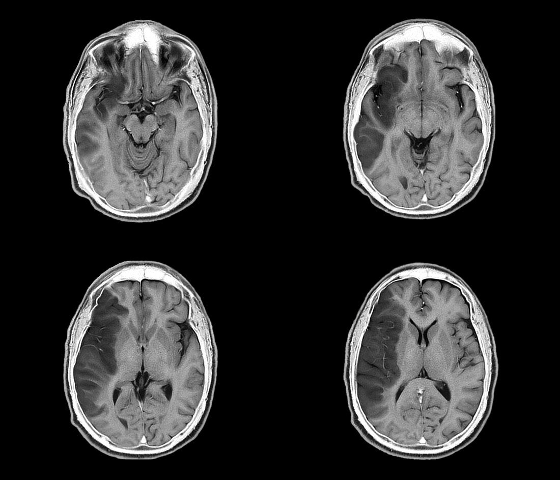
| Stroke. Axial magnetic resonance imaging (MRI) scans through the brain of a 54-year-old woman who has had a stroke that has caused partial left-side hemiplegia (paralysis of one side of the body). These scans were performed 2 hours after the stroke. The damaged area of the brain is at upper left and centre left in each image, on the right-hand side of the brain. This stroke (also called a cerebrovascular accident, CVA) is due to reduced blood supply (ischaemia), probably due to a blockage of an artery. These scans are T2 sequence MRIs, with inversion used to highlight the tissues being examined. The sequence starts at the level of the eyes (top left), with the subsequent scans (left to right and top to bottom) at higher levels in the brain, ending at lower right. | |
| Lizenzart: | Lizenzpflichtig |
| Credit: | Science Photo Library / Zephyr |
| Bildgröße: | 4515 px × 3870 px |
| Modell-Rechte: | nicht erforderlich |
| Eigentums-Rechte: | nicht erforderlich |
| Restrictions: | - |
Preise für dieses Bild ab 15 €
Universitäten & Organisationen
(Informationsmaterial Digital, Informationsmaterial Print, Lehrmaterial Digital etc.)
ab 15 €
Redaktionell
(Bücher, Bücher: Sach- und Fachliteratur, Digitale Medien (redaktionell) etc.)
ab 30 €
Werbung
(Anzeigen, Aussenwerbung, Digitale Medien, Fernsehwerbung, Karten, Werbemittel, Zeitschriften etc.)
ab 55 €
Handelsprodukte
(bedruckte Textilie, Kalender, Postkarte, Grußkarte, Verpackung etc.)
ab 75 €
Pauschalpreise
Rechtepakete für die unbeschränkte Bildnutzung in Print oder Online
ab 495 €
Keywords
- 50er Jahre,
- abnormal,
- ausgeschnitten,
- Ausschnitte,
- axial,
- cerebral,
- CVA,
- Diagnose,
- Einfarbig,
- Erwachsene,
- Frau,
- Fünfziger Jahre,
- geduldig,
- Gehirn,
- Hemiplegie,
- Ischämie,
- Kondition,
- Kopf,
- Krankheit,
- Lähmung,
- Magnetresonanztomografie,
- Medizin,
- medizinisch,
- menschlicher Körper,
- MRT-Untersuchung,
- Neurologie,
- neurologisch,
- Niemand,
- Quartett,
- Querschnitt,
- Reihenfolge,
- Scanner,
- Schlaganfall,
- Schwarz und weiß,
- schwarzer Hintergrund,
- Sektion,
- sektioniert,
- Serie,
- Störung,
- ungesund,
- vaskulär,
- vier,
- Weiblich