Bones of the hip joint, 3D CT scan
Bildnummer 12378941
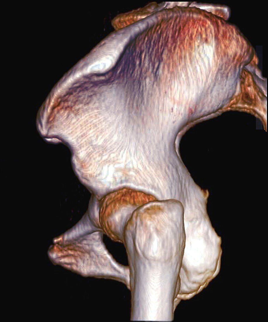
| Bones of the hip joint. Coloured 3D computed tomography (CT) scan of a side view of the left hip joint of a 28-year-old woman. The front of the body is at left. The acetabulum (hip socket) is where the pelvis bones articulate with the head of the femur (thigh bone) to form the hip joint. The main pelvic bones are known as the coxal bones, and consist of the ilium, ischium, and pubis. The pelvic bones visible here are the ilium (wing-shaped, top), as well as a partial view of the ischium and the pubis (lower left). | |
| Lizenzart: | Lizenzpflichtig |
| Credit: | Science Photo Library / Zephyr |
| Bildgröße: | 3812 px × 4585 px |
| Modell-Rechte: | nicht erforderlich |
| Eigentums-Rechte: | nicht erforderlich |
| Restrictions: | - |
Preise für dieses Bild ab 15 €
Universitäten & Organisationen
(Informationsmaterial Digital, Informationsmaterial Print, Lehrmaterial Digital etc.)
ab 15 €
Redaktionell
(Bücher, Bücher: Sach- und Fachliteratur, Digitale Medien (redaktionell) etc.)
ab 30 €
Werbung
(Anzeigen, Aussenwerbung, Digitale Medien, Fernsehwerbung, Karten, Werbemittel, Zeitschriften etc.)
ab 55 €
Handelsprodukte
(bedruckte Textilie, Kalender, Postkarte, Grußkarte, Verpackung etc.)
ab 75 €
Pauschalpreise
Rechtepakete für die unbeschränkte Bildnutzung in Print oder Online
ab 495 €
Keywords
- 20er Jahre,
- 3 dimensional,
- 3-d,
- 3-dimensional,
- 3D,
- Anatomie,
- anatomisch,
- Arthrologie,
- Becken,
- Bein,
- Biologie,
- biologisch,
- Computertomographie,
- CT-Scan,
- Dreidimensional,
- dreißiger Jahre,
- Erwachsene,
- farbig,
- Femur,
- Frau,
- gefärbt,
- Gelenk,
- gesund,
- Hüfte,
- Hüften,
- Joint,
- Knochen,
- menschlicher Körper,
- Niemand,
- normal,
- Osteologie,
- pelvin,
- Profil,
- Scanner,
- schwarzer Hintergrund,
- Seitenansicht,
- seitlich,
- Weiblich