Intestinal tissue, fluorescent light micrograph
Bildnummer 12377244
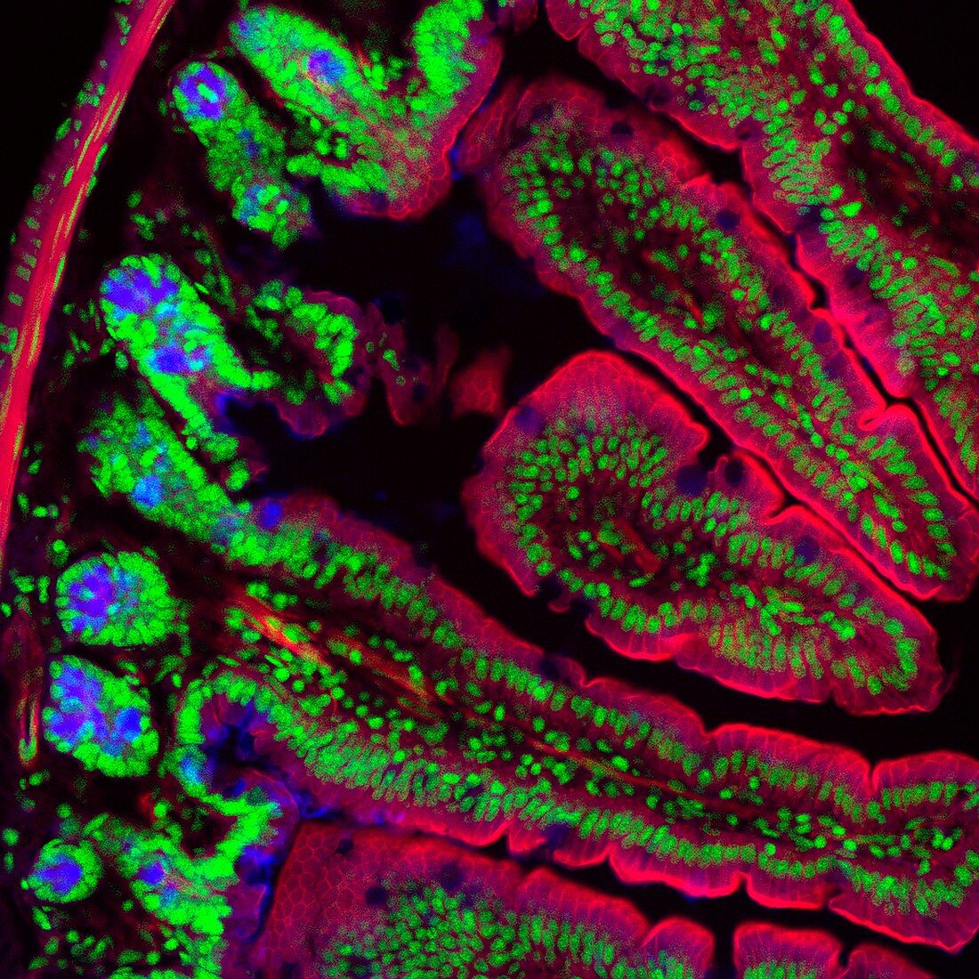
| Intestinal tissue. Confocal fluorescent light micrograph of a section through tissue from the intestines, showing the folded structure and cell proteins. The structures and proteins highlighted are cell nuclei (green), actin (red), and the basement membrane and crypts (blue). This sample is from the intestines of a mouse. | |
| Lizenzart: | Lizenzpflichtig |
| Credit: | Science Photo Library / UNIVERSITY OF ABERDEEN / Kevin Mackenzie |
| Bildgröße: | 2956 px × 2956 px |
| Modell-Rechte: | nicht erforderlich |
| Eigentums-Rechte: | nicht erforderlich |
| Restrictions: | - |
Preise für dieses Bild ab 15 €
Universitäten & Organisationen
(Informationsmaterial Digital, Informationsmaterial Print, Lehrmaterial Digital etc.)
ab 15 €
Redaktionell
(Bücher, Bücher: Sach- und Fachliteratur, Digitale Medien (redaktionell) etc.)
ab 30 €
Werbung
(Anzeigen, Aussenwerbung, Digitale Medien, Fernsehwerbung, Karten, Werbemittel, Zeitschriften etc.)
ab 55 €
Handelsprodukte
(bedruckte Textilie, Kalender, Postkarte, Grußkarte, Verpackung etc.)
ab 75 €
Pauschalpreise
Rechtepakete für die unbeschränkte Bildnutzung in Print oder Online
ab 495 €
Keywords
- Aktin,
- Biologie,
- biologisch,
- Darm,
- Darm-,
- Fluoreszenz,
- fluoreszierend,
- Gedärme,
- gesund,
- Gewebe,
- Histologie,
- histologisch,
- konfokal,
- Lichtmikroskop,
- lichtmikroskopische Aufnahme,
- Maus,
- Niemand,
- normal,
- Proteine,
- Querschnitt,
- Sektion,
- sektioniert,
- Tierkörper,
- Trakt,
- Verdauung,
- Verdauungssystem,
- Zellbilogie,
- Zelle,
- Zellen,
- zellular