Intestinal tissue, TEM
Bildnummer 12377243
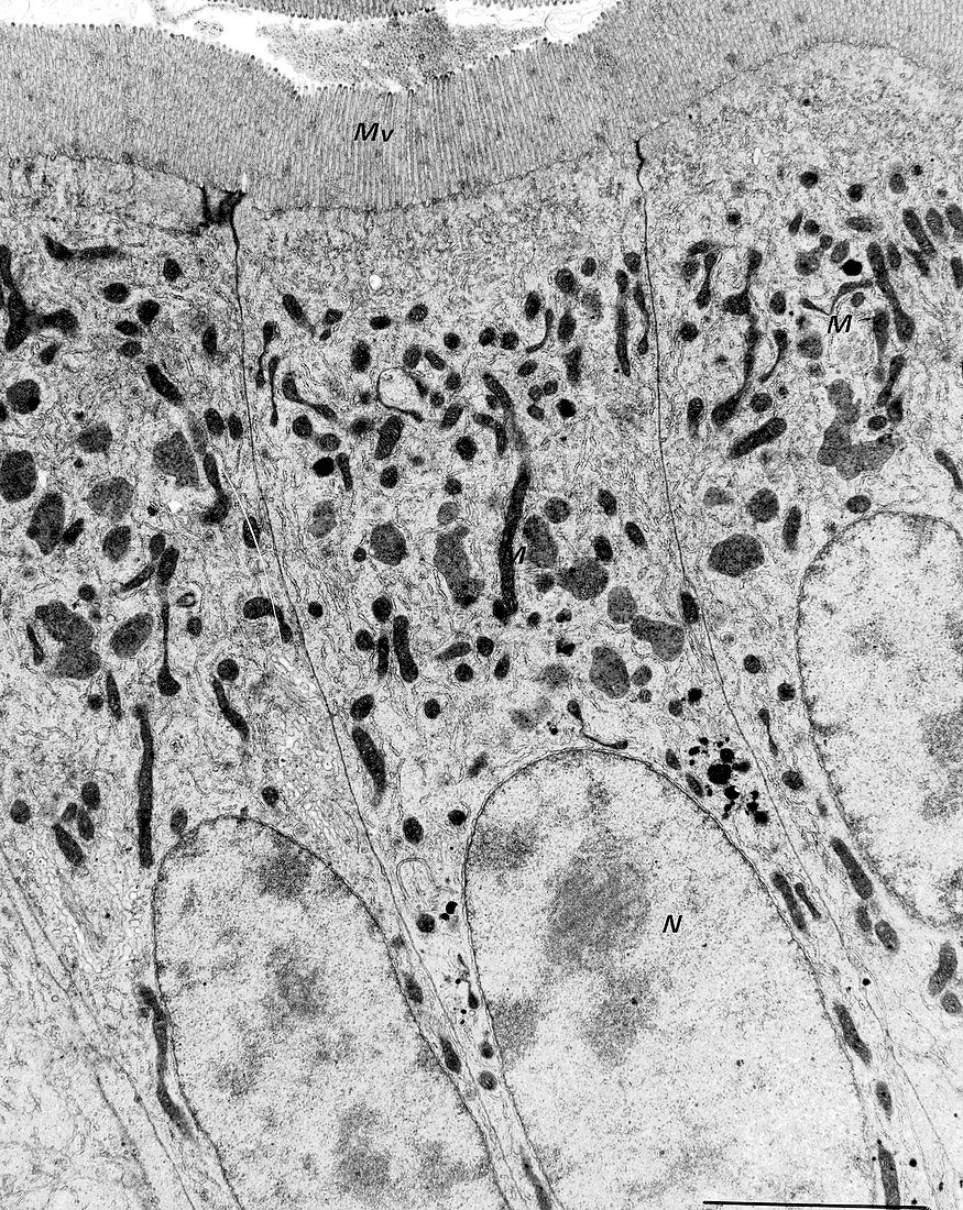
| Intestinal tissue, transmission electron micrograph (TEM). The structures visible here are the cells of the columnar epithelium, with large columnar cells topped by microvilli (top, labelled 'Mv')). The nuclei of the epithelial cells are across bottom (labelled 'N'). At centre are the mucus-secreting areas ('M'). | |
| Lizenzart: | Lizenzpflichtig |
| Credit: | Science Photo Library / UNIVERSITY OF ABERDEEN / Kevin Mackenzie |
| Bildgröße: | 2307 px × 2900 px |
| Modell-Rechte: | nicht erforderlich |
| Eigentums-Rechte: | nicht erforderlich |
| Restrictions: | - |
Preise für dieses Bild ab 15 €
Universitäten & Organisationen
(Informationsmaterial Digital, Informationsmaterial Print, Lehrmaterial Digital etc.)
ab 15 €
Redaktionell
(Bücher, Bücher: Sach- und Fachliteratur, Digitale Medien (redaktionell) etc.)
ab 30 €
Werbung
(Anzeigen, Aussenwerbung, Digitale Medien, Fernsehwerbung, Karten, Werbemittel, Zeitschriften etc.)
ab 55 €
Handelsprodukte
(bedruckte Textilie, Kalender, Postkarte, Grußkarte, Verpackung etc.)
ab 75 €
Pauschalpreise
Rechtepakete für die unbeschränkte Bildnutzung in Print oder Online
ab 495 €
Keywords
- Biologie,
- biologisch,
- Bürstensaum,
- Darm,
- Darm-,
- Einfarbig,
- Gedärme,
- gesund,
- Gewebe,
- Histologie,
- histologisch,
- Kerne,
- menschlicher Körper,
- Mikrovilli,
- Mikrovillus,
- Niemand,
- normal,
- Organelle,
- Organellen,
- Querschnitt,
- Schleim,
- Schleimhaut,
- Schwarz und weiß,
- Sektion,
- sektioniert,
- tem,
- Trakt,
- Transmissionselektronenmikroskop,
- transmissionselektronenmikroskopische Aufnahme,
- Verdauung,
- Verdauungssystem,
- Zellbilogie,
- Zelle,
- Zellen,
- Zellkern,
- zellular