Skeletal bone core surface, SEM
Bildnummer 12377231
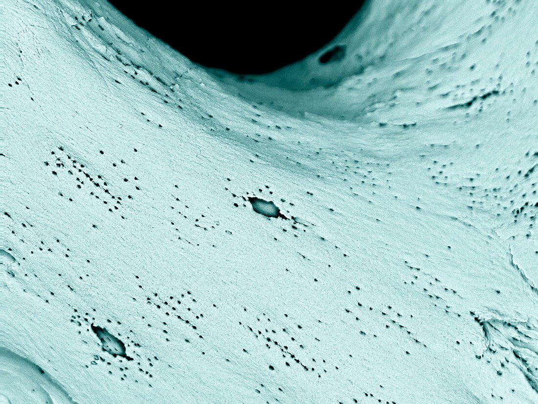
| Skeletal bone core surface. Coloured scanning electron micrograph (SEM) of the surface of bone tissue in a bone core taken from a femur (thigh bone) and cleaned with enzymes to remove all soft tissues. This reveals the underlying bone structure, which includes osteocytes (bone cells) in this close-up view. Imaged using a backscatter electron detector (BSD). | |
| Lizenzart: | Lizenzpflichtig |
| Credit: | Science Photo Library / UNIVERSITY OF ABERDEEN / Kevin Mackenzie |
| Bildgröße: | 3413 px × 2559 px |
| Modell-Rechte: | nicht erforderlich |
| Eigentums-Rechte: | nicht erforderlich |
| Restrictions: | - |
Preise für dieses Bild ab 15 €
Universitäten & Organisationen
(Informationsmaterial Digital, Informationsmaterial Print, Lehrmaterial Digital etc.)
ab 15 €
Redaktionell
(Bücher, Bücher: Sach- und Fachliteratur, Digitale Medien (redaktionell) etc.)
ab 30 €
Werbung
(Anzeigen, Aussenwerbung, Digitale Medien, Fernsehwerbung, Karten, Werbemittel, Zeitschriften etc.)
ab 55 €
Handelsprodukte
(bedruckte Textilie, Kalender, Postkarte, Grußkarte, Verpackung etc.)
ab 75 €
Pauschalpreise
Rechtepakete für die unbeschränkte Bildnutzung in Print oder Online
ab 495 €
Keywords
- Anatomie,
- anatomisch,
- Biologie,
- biologisch,
- Close-up,
- Detail,
- farbig,
- Femur,
- gefärbt,
- gesund,
- Gewebe,
- Knochen,
- menschlicher Körper,
- Mineral,
- Mineralien,
- Niemand,
- normal,
- Oberfläche,
- Oberschenkelknochen,
- Osteologie,
- Osteozyten,
- Rasterelektronenmikroskop,
- rasterelektronenmikroskopische Aufnahme,
- REM,
- Skelettknochen