Thymus, light micrograph
Bildnummer 12360986
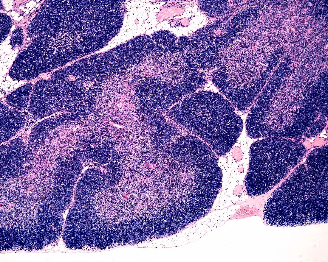
| Low magnification light micrograph showing a young thymus. The organization into lobules is clearly seen. In each lobule, the peripheral cortex appears more stained, due to the high density of T-lymphocyte precursor cells. In the paler centre of each lobule there are many Hassall's corpuscles. Among the lobules, there is a small amount of adipose tissue. Magnification: x36 when printed at 10 centimetres across. | |
| Lizenzart: | Lizenzpflichtig |
| Credit: | Science Photo Library / JOSE CALVO |
| Bildgröße: | 5760 px × 4608 px |
| Modell-Rechte: | nicht erforderlich |
| Eigentums-Rechte: | nicht erforderlich |
| Restrictions: | - |
Preise für dieses Bild ab 15 €
Universitäten & Organisationen
(Informationsmaterial Digital, Informationsmaterial Print, Lehrmaterial Digital etc.)
ab 15 €
Redaktionell
(Bücher, Bücher: Sach- und Fachliteratur, Digitale Medien (redaktionell) etc.)
ab 30 €
Werbung
(Anzeigen, Aussenwerbung, Digitale Medien, Fernsehwerbung, Karten, Werbemittel, Zeitschriften etc.)
ab 55 €
Handelsprodukte
(bedruckte Textilie, Kalender, Postkarte, Grußkarte, Verpackung etc.)
ab 75 €
Pauschalpreise
Rechtepakete für die unbeschränkte Bildnutzung in Print oder Online
ab 495 €
Keywords
- befleckt,
- Biologie,
- biologisch,
- Blutgefäß,
- Cortex,
- Gefäße,
- gesund,
- Histologie,
- histologisch,
- Immunologie,
- immunologisch,
- Immunsystem,
- Lichtmikroskop,
- lichtmikroskopische Aufnahme,
- Lymphozyten,
- Makrophagen,
- Mark,
- Menschliche Biologie,
- menschlicher Körper,
- Mikrofotografie,
- Mikroskopie,
- mikroskopisch,
- Sektion,
- T-Zelle,
- Verfärbung,
- weißes Blutkörperchen,
- Zellbilogie,
- Zytologie,
- Zytologisch