Convoluted tubules, light micrograph
Bildnummer 12360952
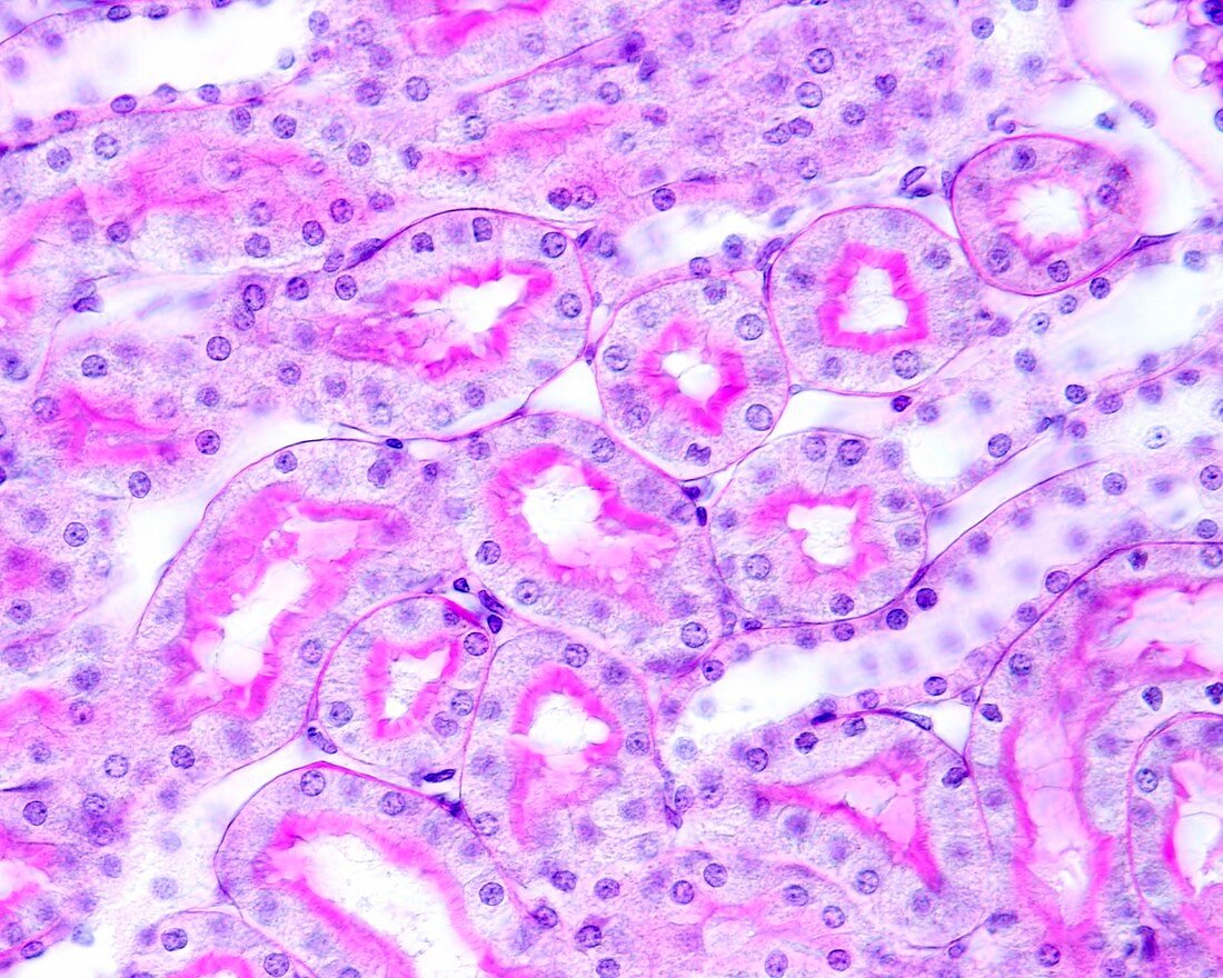
| High magnification light micrograph of a renal cortex stained with the periodic acid method of Schiff (PAS). In the proximal convoluted tubules, the PAS method highlights the basement membrane and the brush border bordering the lumen. There are also several distal convoluted tubules (two longitudinally sectioned on the right edge of the image), with a lower epithelium and lacking a brush border at the luminal edge. Magnification: x360 when printed at 10 centimetres across. | |
| Lizenzart: | Lizenzpflichtig |
| Credit: | Science Photo Library / JOSE CALVO |
| Bildgröße: | 4674 px × 3739 px |
| Modell-Rechte: | nicht erforderlich |
| Eigentums-Rechte: | nicht erforderlich |
| Restrictions: | - |
Preise für dieses Bild ab 15 €
Universitäten & Organisationen
(Informationsmaterial Digital, Informationsmaterial Print, Lehrmaterial Digital etc.)
ab 15 €
Redaktionell
(Bücher, Bücher: Sach- und Fachliteratur, Digitale Medien (redaktionell) etc.)
ab 30 €
Werbung
(Anzeigen, Aussenwerbung, Digitale Medien, Fernsehwerbung, Karten, Werbemittel, Zeitschriften etc.)
ab 55 €
Handelsprodukte
(bedruckte Textilie, Kalender, Postkarte, Grußkarte, Verpackung etc.)
ab 75 €
Pauschalpreise
Rechtepakete für die unbeschränkte Bildnutzung in Print oder Online
ab 495 €
Keywords
- Adventitia,
- Anatomie,
- anatomisch,
- Ausscheidung,
- Bindegewebe,
- Biologie,
- biologisch,
- Blase,
- Blutgefäß,
- Blutgefäße,
- Bürstensaum,
- Epithel,
- epithelial,
- Epithelien,
- Erythrozyt,
- Erythrozyten,
- Gefäß,
- Gefäße,
- gesund,
- Gewebe,
- glomerulär,
- Glomerulus,
- Harnsystem,
- Henle-Schleife,
- Histologie,
- histologisch,
- kapillar,
- Lichtmikroskop,
- lichtmikroskopische Aufnahme,
- Lumen,
- Mark,
- Menschliche Biologie,
- menschlicher Körper,
- Mikrofotografie,
- Mikroskop,
- Mikroskopie,
- mikroskopisch,
- Mikrovilli,
- Mikrovillus,
- nephron,
- Niere,
- Nieren,
- Nieren-,
- Plattenepithel,
- Querschnitt,
- rot,
- rote Blutkörperchen,
- Schleimhaut,
- Submukosa,
- Übergangsepithel,
- Vergrößerung,
- Zelle,
- Zellen