Cancellous bone, light micrographs
Bildnummer 12360950
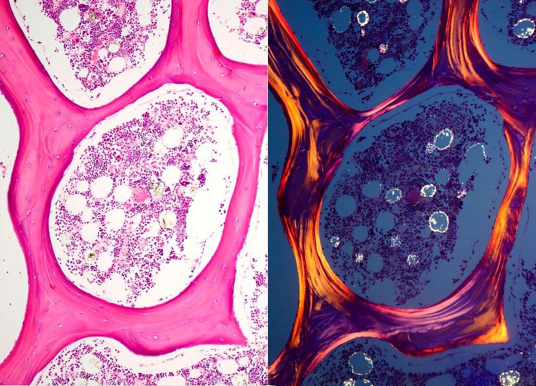
| Comparative light micrographs of cancellous, or spongy, bone stained with haematoxylin and eosin (HE, left) and seen under polarized light (right). With HE, the organization of spongy bone tissue is seen with trabeculae that connect with one another leaving large spaces occupied by bone marrow. With HE, the osteocytes of the trabeculae can be seen. Under polarized light the different orientations of the collagen microfibers that make up each bony lamina lead to alternate bright and dark bands in the bone trabeculae. In the bone marrow some adipocytes show high brightness because their birefringence. Magnification: x90 when printed at 10 centimetres across. | |
| Lizenzart: | Lizenzpflichtig |
| Credit: | Science Photo Library / JOSE CALVO |
| Bildgröße: | 4923 px × 3550 px |
| Modell-Rechte: | nicht erforderlich |
| Eigentums-Rechte: | nicht erforderlich |
| Restrictions: | - |
Preise für dieses Bild ab 15 €
Universitäten & Organisationen
(Informationsmaterial Digital, Informationsmaterial Print, Lehrmaterial Digital etc.)
ab 15 €
Redaktionell
(Bücher, Bücher: Sach- und Fachliteratur, Digitale Medien (redaktionell) etc.)
ab 30 €
Werbung
(Anzeigen, Aussenwerbung, Digitale Medien, Fernsehwerbung, Karten, Werbemittel, Zeitschriften etc.)
ab 55 €
Handelsprodukte
(bedruckte Textilie, Kalender, Postkarte, Grußkarte, Verpackung etc.)
ab 75 €
Pauschalpreise
Rechtepakete für die unbeschränkte Bildnutzung in Print oder Online
ab 495 €
Keywords
- Anatomie,
- anatomisch,
- befleckt,
- Biologie,
- biologisch,
- gesund,
- Gewebe,
- Histologie,
- histologisch,
- Kalzium,
- Knochen,
- Kollagen,
- Lichtmikroskop,
- lichtmikroskopische Aufnahme,
- Matrix,
- menschlicher Körper,
- Mikroskopie,
- Mineral,
- Osteon,
- Osteozyten,
- Sektion,
- sektioniert,
- Skelettknochen,
- Zelle,
- Zellen,
- Zytologie,
- Zytologisch