Bone growth plate, light micrograph
Bildnummer 12360928
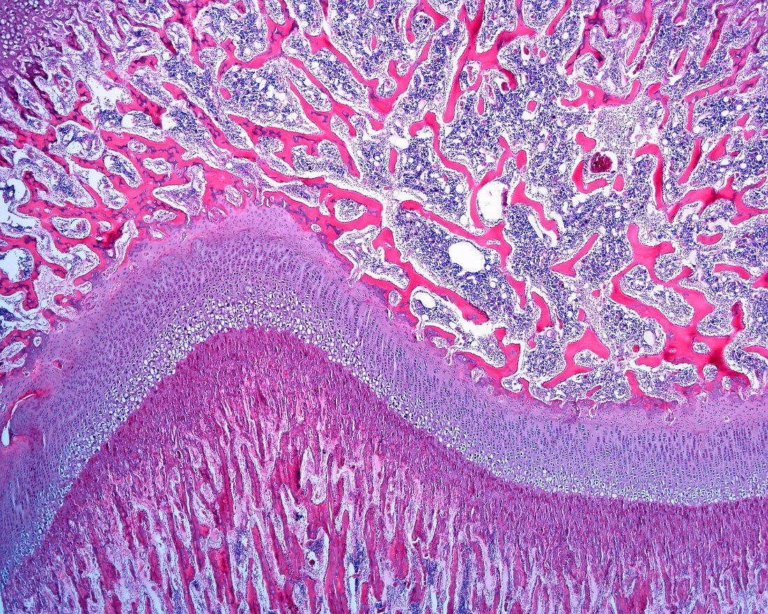
| Low magnification light micrograph of the epiphyseal growth plate of a developing long bone. In the upper part of the image, above the epiphyseal cartilage, is the epiphysis, in the process of ossification. It consists of mixed bone trabeculae with a calcified cartilage centre (very thin) and a thick lining of bone tissue. The epiphyseal cartilage appears curved in the image. Although the magnification is very low, a clear area (corresponding to hypertrophic cartilage) is well distinguished. Underneath, there is a compact stripe (the forward front of the centre of ossification) and, further down, a zone of mixed trabeculae. Magnification: x18 when printed at 10 centimetres across. | |
| Lizenzart: | Lizenzpflichtig |
| Credit: | Science Photo Library / JOSE CALVO |
| Bildgröße: | 5760 px × 4608 px |
| Modell-Rechte: | nicht erforderlich |
| Eigentums-Rechte: | nicht erforderlich |
| Restrictions: | - |
Preise für dieses Bild ab 15 €
Universitäten & Organisationen
(Informationsmaterial Digital, Informationsmaterial Print, Lehrmaterial Digital etc.)
ab 15 €
Redaktionell
(Bücher, Bücher: Sach- und Fachliteratur, Digitale Medien (redaktionell) etc.)
ab 30 €
Werbung
(Anzeigen, Aussenwerbung, Digitale Medien, Fernsehwerbung, Karten, Werbemittel, Zeitschriften etc.)
ab 55 €
Handelsprodukte
(bedruckte Textilie, Kalender, Postkarte, Grußkarte, Verpackung etc.)
ab 75 €
Pauschalpreise
Rechtepakete für die unbeschränkte Bildnutzung in Print oder Online
ab 495 €