Mixed tubuloacinar gland, light micrograph
Bildnummer 12360926
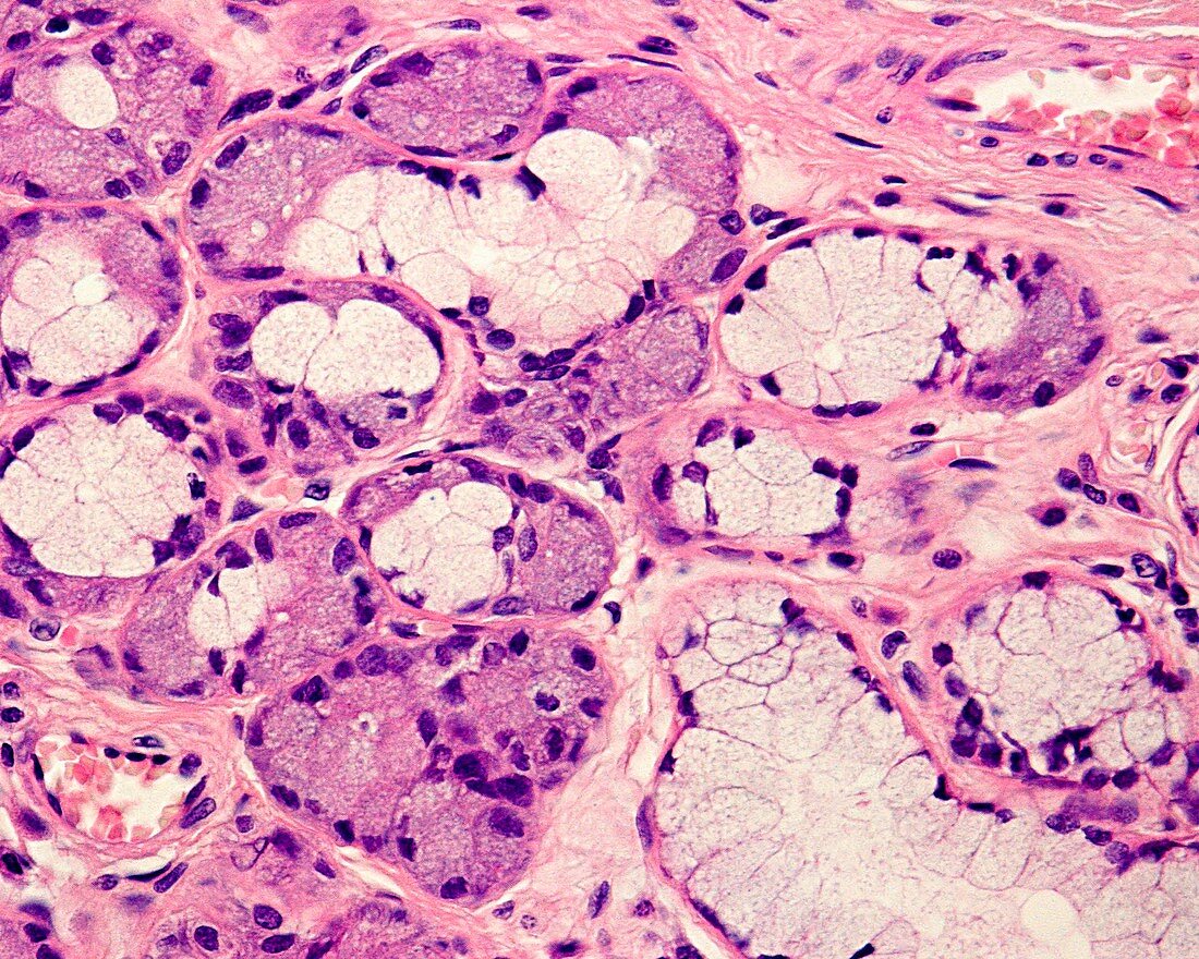
| Salivary gland. High magnification light micrograph showing several mixed tubulo-acini. Note the location of the serous cells in the bottom of the acinus, the cell clusters are crescent-shaped, which gives rise to their name - Giannuzzi's demilunes. Magnification: x360 when printed at 10 centimetres across. | |
| Lizenzart: | Lizenzpflichtig |
| Credit: | Science Photo Library / JOSE CALVO |
| Bildgröße: | 4674 px × 3739 px |
| Modell-Rechte: | nicht erforderlich |
| Eigentums-Rechte: | nicht erforderlich |
| Restrictions: | - |
Preise für dieses Bild ab 15 €
Universitäten & Organisationen
(Informationsmaterial Digital, Informationsmaterial Print, Lehrmaterial Digital etc.)
ab 15 €
Redaktionell
(Bücher, Bücher: Sach- und Fachliteratur, Digitale Medien (redaktionell) etc.)
ab 30 €
Werbung
(Anzeigen, Aussenwerbung, Digitale Medien, Fernsehwerbung, Karten, Werbemittel, Zeitschriften etc.)
ab 55 €
Handelsprodukte
(bedruckte Textilie, Kalender, Postkarte, Grußkarte, Verpackung etc.)
ab 75 €
Pauschalpreise
Rechtepakete für die unbeschränkte Bildnutzung in Print oder Online
ab 495 €
Keywords
- Biologie,
- biologisch,
- Drüse,
- exokrine Drüse,
- Fotografie,
- Gang,
- gastrointestinal,
- gesund,
- Histologie,
- histologisch,
- lichtmikroskopische Aufnahme,
- Magen-Darm-Trakt,
- Mensch,
- Menschliche Biologie,
- menschlicher Körper,
- Mikrofotografie,
- Mikroskopie,
- mikroskopisch,
- Querschnitt,
- Schleim,
- sekretorisch,
- Verdauungs-,
- Verdauungssystem,
- Verdauungstrakt