Pituitary gland, light micrograph
Bildnummer 12360909
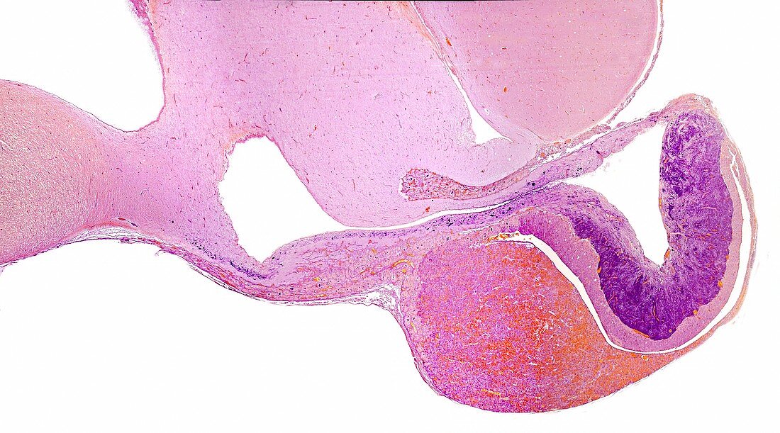
| Very low magnification light micrograph showing a panoramic view of a cat pituitary gland in a sagittal section. From left to right, the hypothalamus, the pituitary stalk (almost horizontal), the anterior lobe of the adenohypophysis, the Rathke's cleft (which in this species remains throughout adult life), the intermediate lobe and the posterior lobe (with its neurosecretion stained in magenta). Gabe's paraldehyde-fuchsin method. Magnification: x18 when printed at 10 centimetres across. | |
| Lizenzart: | Lizenzpflichtig |
| Credit: | Science Photo Library / JOSE CALVO |
| Bildgröße: | 5607 px × 3116 px |
| Modell-Rechte: | nicht erforderlich |
| Eigentums-Rechte: | nicht erforderlich |
| Restrictions: | - |
Preise für dieses Bild ab 15 €
Universitäten & Organisationen
(Informationsmaterial Digital, Informationsmaterial Print, Lehrmaterial Digital etc.)
ab 15 €
Redaktionell
(Bücher, Bücher: Sach- und Fachliteratur, Digitale Medien (redaktionell) etc.)
ab 30 €
Werbung
(Anzeigen, Aussenwerbung, Digitale Medien, Fernsehwerbung, Karten, Werbemittel, Zeitschriften etc.)
ab 55 €
Handelsprodukte
(bedruckte Textilie, Kalender, Postkarte, Grußkarte, Verpackung etc.)
ab 75 €
Pauschalpreise
Rechtepakete für die unbeschränkte Bildnutzung in Print oder Online
ab 495 €