Human heart atrium, light micrograph
Bildnummer 12360900
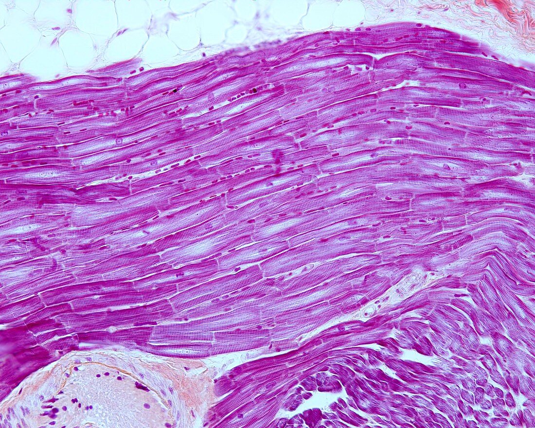
| Light micrograph of a human atrial myocardium stained with Mallory's phosphotungstic hematoxylin. A group of longitudinally sectioned cardiac myocytes, shows cross-striation. Nuclei are located in the centre of the cell and intercalated discs in the intercellular contacts. The rows of small dark spots located between the myocytes are red blood cells. Magnification: x360 when printed at 10 centimetres across. | |
| Lizenzart: | Lizenzpflichtig |
| Credit: | Science Photo Library / JOSE CALVO |
| Bildgröße: | 4674 px × 3739 px |
| Modell-Rechte: | nicht erforderlich |
| Eigentums-Rechte: | nicht erforderlich |
| Restrictions: | - |
Preise für dieses Bild ab 15 €
Universitäten & Organisationen
(Informationsmaterial Digital, Informationsmaterial Print, Lehrmaterial Digital etc.)
ab 15 €
Redaktionell
(Bücher, Bücher: Sach- und Fachliteratur, Digitale Medien (redaktionell) etc.)
ab 30 €
Werbung
(Anzeigen, Aussenwerbung, Digitale Medien, Fernsehwerbung, Karten, Werbemittel, Zeitschriften etc.)
ab 55 €
Handelsprodukte
(bedruckte Textilie, Kalender, Postkarte, Grußkarte, Verpackung etc.)
ab 75 €
Pauschalpreise
Rechtepakete für die unbeschränkte Bildnutzung in Print oder Online
ab 495 €
Keywords
- Anatomie,
- anatomisch,
- arteriell,
- Biologie,
- biologisch,
- Blut,
- Erythrozyt,
- Gefäßsystem,
- gesund,
- Gesundheitswesen,
- Gewebe,
- Hämatoxylin,
- Herz,
- Histologie,
- histologisch,
- kardiovaskular,
- Kreislauf,
- Lichtmikroskop,
- lichtmikroskopische Aufnahme,
- Lumen,
- Medizin,
- medizinisch,
- Mensch,
- menschlicher Körper,
- Mikrofotografie,
- Mikrographie,
- Mikrophotographie,
- Mikroskopie,
- Muskel,
- Myokard,
- Myozyten,
- normal,
- Sektion,
- sektioniert,
- Verfärbung,
- Zelle,
- Zellen,
- zellular